Download
1 / 10
160 likes | 613 Views
Cell types. Extracellular matrix. Ground substance. Fibers • Collagen fiber • Elastic fiber • Reticular fiber. Macrophage. Fibroblast. Lymphocyte. Fat cell. Capillary. Mast cell. Neutrophil. Figure 4.7. Connective Tissue: Embryonic. Mesenchyme—embryonic connective tissue

E N D
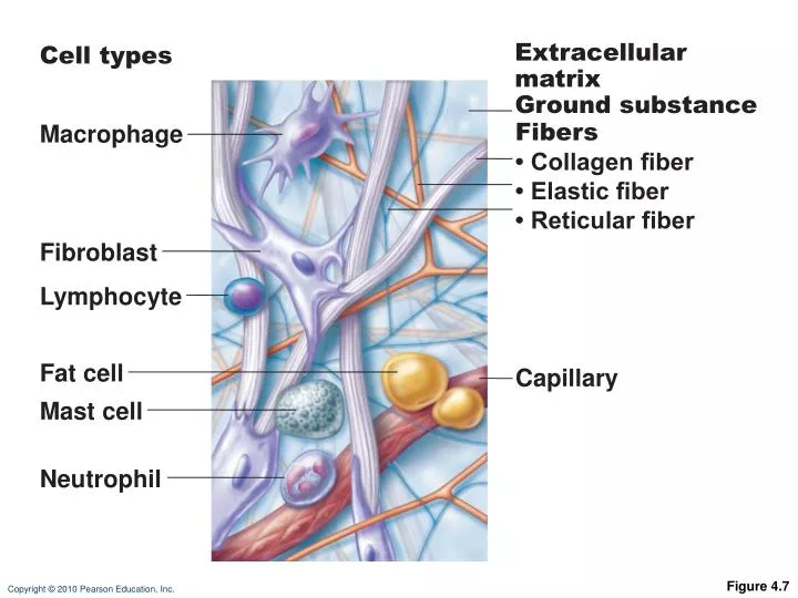
Cell types Extracellular matrix Ground substance Fibers • Collagen fiber • Elastic fiber • Reticular fiber Macrophage Fibroblast Lymphocyte Fat cell Capillary Mast cell Neutrophil Figure 4.7
Connective Tissue: Embryonic • Mesenchyme—embryonic connective tissue • Gives rise to all other connective tissues • Gel-like ground substance with fibers and star-shaped mesenchymal cells
Overview of Connective Tissues • For each of the following examples of connective tissue, note: • Description • Function • Location
Teratomas are made up of a variety of parenchymal cell types representing more than 1 germ layer. The most common location is sacrococcygeal (57%). Because some arise from totipotential cells, they are encountered commonly in the gonads (29%). By far, the most common gonadal location is the ovary. Cells differentiate along various germ lines, essentially recapitulating any tissue of the body. Examples include hair, teeth, fat, skin, muscle, and endocrine tissue
Epithelial Membranes • Mucous membranes • Mucosae • Line body cavities open to the exterior (e.g., digestive and respiratory tracts)
Mucosa of nasal cavity Mucosa of mouth Esophagus lining Mucosa of lung bronchi (b) Mucous membranes line body cavitiesopen to the exterior. Figure 4.11b
Epithelial Membranes • Serous Membranes • Serosae—membranes (mesothelium + areolar tissue) in a closed ventral body cavity • Parietal serosae line internal body walls • Visceral serosae cover internal organs
Parietal peritoneum Parietal pleura Visceral pleura Visceral peritoneum Parietal pericardium Visceral pericardium (c) Serous membranes line body cavitiesclosed to the exterior. Figure 4.11c