Download
1 / 49
500 likes | 641 Views
Cells and Tissue. Chapter 3. Cells . Made up of 4 elements C,O,H,N smaller elements Ca2+, Fe 60% water, essential for life Bathed in dilute saltwater solution = interstitial fluid Exchanges between blood and cells through interstitial fluid.

E N D
Cells and Tissue Chapter 3
Cells • Made up of 4 elements • C,O,H,N • smaller elements Ca2+, Fe • 60% water, essential for life • Bathed in dilute saltwater solution = interstitial fluid • Exchanges between blood and cells through interstitial fluid http://www.guardian.co.uk/science/2009/jan/08/stem-cells-bone-marrow-heart-attack
Cells • Range in length - 2μm-1+m • Structure reflects function • Disc • Threadlike extensions • Toothpick shaped • Function varies • Phagocytosis • Synthesize hormones • Cleanse blood http://www.wereyouwondering.com/what-is-the-smallest-cell-in-the-human-body/ http://www.ngfn.de/englisch/glossar443.htm
Anatomy of Generalized Cell • No 2 cell types the same, some functions common to all • Nucleus, Cytoplasm, Plasma membrane Figure 3.4
Anatomy of Generalized CellNucleus • Control center • Gene-containing • Genetic material DNA • Instructions for building proteins • Necessary for cell reproduction • Usually round or oval, but conforms to shape of cell • Nuclear Envelope • Nucleoli • Chromatin
NucleusNuclear Envelope • Nucleus bound by double membrane • Space between layers filled with fluid • Nuclear pores – points where two layers fuse • Selectively permeable, but large pores • Encloses nucleoplasm – suspends nuclear elements
NucleusNucleoli • Small, round body • Site for ribosome assembly • Ribosomes migrate to cytoplasm, site of protein synthesis
NucleusChromatin • When cell not dividing, DNA associated with proteins • Loose coiling of DNA and proteins called chromatin • When cell is dividing, DNA and proteins condense to form chromosomes
Plasma Membrane • Separates cell from surroundings • Lipid bilayer (tail to tail) – protein molecules float • Phospholipids • Cholesterol – keeps membrane fluid Figure 3.2
Plasma MembranePhospholipid bilayer • Semi-permeable • Heads of phospholipids hydrophilic – attracted to water in extra and intercellular fluid • Tails are hydrophobic – avoid water and line up in center • Structure keeps polar molecules from passing through membrane
Plasma MembraneProteins in membrane • Specialized function of membrane • Receptors • Enzymes • Anchors • Transport • Channels • Glycoproteins – sugars attached to proteins • Surface of cell sticky, fuzzy = glycocalyx • Determine blood type, receptors bacteria, viruses, toxins bind to • Cell to cell interaction • See changes here when cell becomes cancerous
Plasma MembraneSpecializations • Microvilli – fingerlike projections that increase SA (intestines) for absorption, speed • Membrane junctions • Tight junctions – impermeable junctions, bind cells together, small intestine digestive enzymes into blood • Gap Junctions – Communication, chemical signals or nutrients pass directly. Neighbors connected through connexons – hollow proteins –heart and embryonic cells • Desmosomes – Anchoring, prevents cells prone to stress from separating - skin • Buttons of p.m. with fine protein filaments. Thick filaments from plaques on one to side to other Figure 3.3
Cytoplasm • Outside nucleus, inside PM • Factory area • 3 major elements • Cytosol • Organelles • Inclusions
CytoplasmCytosol • Semitransparent fluid suspends other things • Mostly water • Nutrients • Other solutes
CytoplasmOrganelles and Inclusions • Organelles • Metabolic machinery • Specialized functions • Inclusions • Chemical substances that vary depending on cell type • Most stored nutrients or cell products • Lipids-fat cells • Glycogen – liver & muscle cells • Pigments (melanin) – skin & hair
OrganellesMitochondria • Double membrane = two PM, side by side • OM smooth • IM has cristae • Enzymes dissolved in fluid w/in mitochondria and in cristae catalyze rxns Oxygen used to break down food • Energy converted to ATP and heat • Powerhouse of cell • Liver and muscle have tons of mitochondria, unfertilized egg has only a few – WHY? http://academic.brooklyn.cuny.edu/biology/bio4fv/page/mito.htm
OrganellesRibosomes • Bilobed, dark bodies • rRNA and proteins • Site of protein synthesis • Float free or attached to RER – synthesize different proteins
OrganellesEndoplasmic Reticulum • System of fluid filled cisterns • Connected to NM • Network for carrying substances (mostly proteins) • Two types: • RER • SER https://illnessesanimalsplants.wikispaces.com/Endoplasmic+Reticulum?f=print
OrganellesEndoplasmic Reticulum - RER • Ribosomes • Membrane factory – building material for membranes synthsized • Proteins made enter cistern, proper conformation, transport vesicle • Abundant is cells that export protein products - pancreas
OrganellesEndoplasmic Reticulum - SER • NO protein synthesis • Lipid synthesis – cholesterol and fat synthesis and breakdown • Detoxification of drugs and pesticides • Common in liver and glands that produce steroid-based hormones
OrganellesGolgi Apparatus • Flattened membranous sacs • Close to nucleus, traffic director to proteins • Modifies and packages proteins from RER depending on function • Proteins tagged for excretion build up in secretory vesicles that pinch off, to PM • Mucus • Digestive enzymes of pancreas • Proteins and phospholipids for PM • Packages digestive enzymes in lysosomes – remain in cell Figure 3.6
OrganellesLysosomes and Peroxisomes • Lysosomes – bags containing digestive enzymes • Demolition sites – digest unused or old organelles, invaders • Abundant in phagocytes • Peroxisomes – Oxidase enzymes, use O2 to detoxify harmful substances – alcohol • Common in kidney cells - detoxification • Disarm free radicals – highly reactive chemicals with unpaired electrons, damage proteins and nucleic acids • Byproduct of metabolism • Convert free radicals to H2O2, Catalase converts to H2O
OrganellesCytoskeleton • Protein network in cytoplasm • Shape, supports organelles, machinery for intracellular transport • 3 types • Microtubules • Overall shape and distribution of organelles • Microfilaments • Cell motility and changes in shape • Intermediate Filaments • Rope-like, desmosomes, resist pulling forces
OrganellesCentrioles • Paired • Rod shaped close to nucleus • In mitosis, direct formation of mitotic spindle • Cilia – projections on some cells, move substances along cell surface – respiratory cells • Flagella – Longer than cilia, motility - sperm
Cell DiversityCells that Connect Body Parts • Erythrocyte • Carries oxygen in bloodstream • Disc shaped • No organelles http://www.becomehealthynow.com/body/cell/erythrocytes.htm
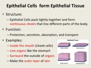
Cell DiversityCells that cover and line body organs • Epithelial Cell • Pack together in sheets • Abundant intermediate filaments – WHY? http://www.covingtoninnovations.com/michael/blog/0707/index.html
Cell DiversityCells that move organs and body parts • Skeletal Muscle and Smooth Muscle • Elongated and filled with contractile filaments • Shorten quickly to move bones or change shape of organ Figure 3.8c
Cell DiversityCells that store nutrients • Spherical in shape • Lipid droplet in cytoplasm Figure 3.8d
Cell DiversityCells that fight disease • Macrophage • Phagocytic • Extends pseudopods to crawl through tissue • Many lysosomes Figure 3.8e
Cell Diversity: Cells that gather info and control functions of the body • Nerve cell (neurons) • Long processes to receive info and transmit to other parts of body • Extensive PM and RER Figure 3.8f
Cell DiversityCells of reproduction • Sperm (male)- long, streamline, built to swim • Oocyte (female) – largest cell in body • Contains several copies of organelles for fertilization Figure 3.8g
Membrane Transport Review solutions, solvents, and solutes! Water is body’s main solvent Solutes are in smaller amounts, so tiny don’t settle out.
Membrane Transport Terms • Intracellular fluid – nucleoplasm and cytosol, sol’n containing gases (O2, CO2), nutrients, salts dissolved in water. • Interstitial fluid – fluid bathes exterior of cells, nutrients (aa, sugars, fatty acids, vitamins), hormones, neurotransmitters, salts, waste. Cells takes what it needs • Selective permeability – some things my pass through, things kept in and out of cell
Membrane TransportPassive Transport: Diffusion • Passive Transport – substances move across membrane w/o energy input from cell • Diffusion – movement of molecule from high concentration to low concentration • Think of crowded elevator or classroom • Saves cells energy • PM is barrier – What can pass? • Small enough to pass through pores • Dissolve in fatty portion of PM (nonpolar) • Assisted by membrane carrier
Types of DiffusionSimple Diffusion • Simple diffusion – unassisted diffusion of solutes through membrane • Lipid-soluble – fats, fat-soluble vitamins, O2, CO2 • Small enough to pass through pores – some ions Cl- Figure 3.10a
Types of DiffusionFacilitated Diffusion Figure 3.10b • Facilitated Diffusion – lipid-insoluble and too large to pass membrane • Proteins channel or carrier molecule transport across • Follows law of diffusion Figure 3.10c
Types of DiffusionOsmosis • Osmosis – diffusion of H2O across membrane • H2O polar, repelled by PM, used aquaporins (proteins in membrane) • Occurs constantly • Glucose and O2 move into cells – WHY? • CO2 and waste moving out – WHY? Figure 3.10d
Membrane TransportPassive Transport - Filtration • Water and solutes FORCED through PM by hydrostatic pressure (blood) • Passive process, but gradient is pressure gradient (higher pressure to lower pressure) • Not selective • Usually occurs in capillaries – • Example – kidney filtration
Membrane TransportActive Transport • Uses ATP • Solute pumps – transport aa, some sugars, most ions • AGAINST GRADIENT • Transports specific substance, way for cell to be selective • Sodium-potassium pump – transmission of nerve impulses Figure 3.11
Membrane TransportActive Transport – Vesicular Transport Some substance can’t get across PM via passive or active transport Uses ATP Moves substance across w/o passing through PM
Vesicular Transport Exocytosis • Moves substance out of cell • How cells secretes hormones, mucus, other products or waste • Packaged by Golgi in vesicles • Migrates to PM and fuses Figure 3.12
Vesicular TransportEndocytosis • Uses ATP • Takes in or engulfs extracellular substances • Moves to cytoplasm binds to lysosome • Pinocytosis – imp. in cells of absorption, but all cells • Phagocytosis – phagocytic cells • Receptor-mediated endocytosis – very selective – enzymes, some hormones, cholesterol Figure 3.13
Cell Division • Cell life cycle – ∆ from formation to division • Two periods • Interphase – growth and normal metabolic activities • DNA is replicated exactly prior to cell division • Semiconservative process – old strand acts as template • Two new complimentary strands • Cell Division – reproduction of cell • Mitosis – division of the nucleus • Cytokinesis – division of cytoplasm
DNA Replication Figure 3.14
Cell DivisionMitosis: Two daughter nuclei with exact same DNA as mother nucleus • Prophase • Chromatin coils and forms chromosomes • DNA duplicated, chromosome = 2 chromatids • Chromatids held together by centromere • Centrioles separate and mitotic spindle forms – transport later • Nuclear membrane and nucleoli broken down Figure 3.15 (1 of 8
Mitosis - Metaphase • Chromosomes line up on metaphase plate Figure 3.15 (6 of 8 )
Mitosis - Anaphase • Chromatids split • Chromosomes pulled to opposite side of cell • Spindles and centromere Figure 3.15 (7 of 8) )
Mitosis – Telophase and Cytokinesis • Telophase • Chromosomes uncoil • Spindles breakdown • NM and nucleoli reform • Cytokinesis • Begins in anaphase, completes in Telophase • Cleavage furrow – microfilaments • Two smaller cells that will grow Figure 3.15 (8 of 8)