Download
1 / 28
330 likes | 715 Views
White Blood Cells Morphology and Counts. Clinical Pathology. Granulocytes. Neutrophils Basophils Eosinophils Produced predominately in the bone marrow. Capable of mitotic division up to the myelocyte stage. Responds to an increased demand infection

E N D
White Blood CellsMorphology and Counts Clinical Pathology
Granulocytes • Neutrophils • Basophils • Eosinophils • Produced predominately in the bone marrow. • Capable of mitotic division up to the myelocyte stage. • Responds to an increased demand • infection • Takes 3-5 days to influence peripheral numbers.
Storage-Maturation Compartment • Cells mature into metamyelocytes→ band cells→ Segmented cells. • 80% of granulocytes are found in the bone marrow of healthy animals. • These cells are released from bone marrow with the oldest (segmented) to increased peripheral need. • In less than two days, the bone marrow can respond. • Dogs have the largest storage pool (5 days) whereas bovines have limited storage.
Functions of Leukocytes • Granulocytes: • Characterized by the presence of cytoplasmic granules • Function of these cells occurs in the tissues, not in the bloodstream. • These cells do not recirculate
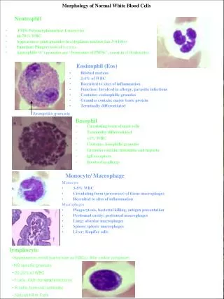
Neutrophils • Primary functions: • Phagocytosis and killing of microbes. • Usually circulate for 10 hours before migrating into the tissues. • Ingest material and eventual bacterial killing.
Eosinophils • Inhibit chemical mediators such as histamine and serotonin which are released during allergic (hypersensitivity reactions). • Have phagocytic and bacteriocidal properties similar to neutrophils but not as effective. • Have parasiticidal properties. • Animals with heartworms may have hig numbers of eosinophils.
Basophils • Secrete mediators of inflammation (histamine) associated with hypersensitivity reactions. • This release occurs when antigens complex with IgE is located on the cellular surface.
Monocytes • Differentiate into the cells of the mononuclear/phagocyte system present in most tissues. • Become macrophages once they migrate into the tissues. • Capable of multiplying within the tissues. • Can survive for long periods of time
Monocyte Functions: • Phagocytosis and digestion of particulate material, bacteria, and dead cells. • Macrophages are less responsive to bacterial infections than neutrophils but are more effective against fungal infections. • Synthesis and release of substances invloved in inflammation and immune response. • Expressions of immune response by presenting antigens to T-lymphocytes. • Serve as a major source of colony stimulating factors and cytokines involved in hemtopoiesis. • Cause bone marrow to produce more granulocytes.
Lymphocytes • Distributed in lymphoid tissue to include lymph nodes, spleen, thymus, tonsils, bone marrow, and blood. • Capable of division. • Recirculate in the blood. • Functions: • B cells: turn into plasma cells which secrete immunoglobulins. Usually stayin the lymphoid tissue. • T cells: transform into effector cells that produce lymphokines which function in mediation of cellular immunity.
Normal morphology • Neutrophils: multiple nuclear lobules separated by constrictions. • Band granulocytes: band (horseshoe) shaped nuclei. • Eosinophil: Lobulated nucleus and cytoplasm containing reddish pink granules. • Basophil: Lobulated nucleus and purple-blue (Basophilic) granules.
Neutrophil compartments in Peripheral blood • Marginated neutrophil pool: during any moment of time, some neutrophils are loosely adhered to the vessel wall. These cells are not sampled when blood is drawn from non-stressed animals. • Circulating neutrophil pool: neutrophils moving with RBC’s and fluid. Are sampled pool of cells. • When an animal is stressed- marginated pool becomes circulating pool. Called stress leukogram.
Response to Inflammation • Inflammatory cells at sites of inflammation release substances (cytokines, interleukins) into the blood to attract neutrophils. • Segmented neutrophils are released from the mature storage compartment in the bone marrow. • Increase in peripheral numbers (measurable in 2 days). • If sudden demand for neutrophils depletes the storage compartment of segmented neutrophils, then band cells are released. • With continued depletion, cells in bone marrow begin to divide. • When source of inflammation is removed, demand for neutrophils decrease, and production slows down.
Immature Neutrophils in Circulation • Immature neutrophils in circulation (left shift)- defined as the release of immature neutrophils (usually bands) into the circulation to meet tissue demand. • The appearance of immature neutrophils is termed “Left shift”. • Regenerative left shift: the absolute number of neutrophils in circulation is increased. The bone marrow has increased the neutrophil release. • Degenerative left shift: the nummber of neutrophils has decreased. This is a poor diagnostic sign.
Cattle exception: • Adult cattle have a relatively low absolute number of neutrophils in circulation and have a small marrow storage pool. • A degenerative left shift is typical of the acute inflammatory response in cattle.
Some terminology for morphology • -penia: decreased number of cells in the blood (Neutropenia, lymphopenia). • -philia or –cytosis: increased number of cells in the blood (neutrophilia, lymphocytosis). • Left shift: increased numbers of immature neutrophils in the blood. • Leukemia: neoplastic cells in the blood or bone marrow. • Leukemoid response: marked leukocytosis (>50,000/ul) usually a result of inflammatory disease. • Lymphoproliferative disorders: conditions in lymphocytes or plasma cells proliferate abnormally. • Myeloproliferative disorders: a group of bone marrow disorders, usually neoplastic, which stems from on of the bone marrow cell lines.
Neutrophilia • Not always due to infection. • Stress leukogram: endogenous or exogenous corticosteroids. • Characterized by neutrophilia without a left shift, monocytosis, lymphopenia, and eosionopenia. • Caused by a shift from the marginal to the circulating pool. • Physiological luekocytosis: transiet condition caused by excitement, epinephrine release and splenic contraction. • Neutrophilia without left shift and normal or increased lymphocyte number. • Occurs more commonly in the cat than the dog.
Inflammatory Leukogram • Increased bone marrow proliferation, shift from storage pool to blood. • Mild inflammation yields a leukocyte response similar to stress. • Purulent reaction: neutrophils with a left shift. • Intense response with left shift • Can confuse immature cells of myeloid series with neoplasia (leukemia) • Degenerative shift usually is due to extreme migration of cells into tissues and/or detrimental side effects of toxins.
Toxic Neutrophils • Characterized by ctyoplasm basophilia, Dohle bodies, toxic granulation, and/or foamy cytoplasm. • Cells have decreased functional abilities. • Animal with toxic, degenerative shift may be compromised by lack of adequate cell number and decrease ability of cells to function.
Dohle Bodies • Blue cytoplasmic inclusions. • Low numbers may be found in healthy cats. • Indicates toxicity in other species.
Neutrophil Hypersegmentation • Neutrophils with five or more nuclear lobes. • Normal aging change of neutrophils which normally occurs in the tissues, not bloodstream. • May occur with excessive levels of corticosteroids from administration or hyperadrenocorticism. • Chronic inflammation. • Artifactual change in the blood that sits for a period of time.
Pelger-Huet Anomaly • Hyposegemented neutrophils that function normally. • Hereditary disorder; failure of the nucleus in mature cells to undergo segmentation.
Lymphocytosis • Physiologic: due to epinephrine release. • Common in chronic inflammation and chronic antigenic stimulation. • Later stages of resolving infections. • Neoplastic lymphocytosis such as leukemia and lymphosarcoma
Lymphopenia • Common finding on CBC • Associated with stress • Immunosuppressive therapy • Immunodeficiency syndrome • Viral infections such as parvo.
Reactive Lymphocytes • Characteristic changes in the morphology of lymphocyte that has been stimulated to produce antibody (B cells) or lymphokines (T cells). • Morphological changes: increased cytoplasm, increased basophilia of cytoplasm, increased aggregation of chromatin, and may lack perinuclear clear zone.