Download
1 / 36
360 likes | 640 Views
Greg Haider. Diagnoses of Cardiac Arrhythmias Through Electrocardiographs. Self Tutorial. To The Index. Index. Importance of EKG Interpretation Review of the Basics The Motor of the Cardiovascular System The EKG process 12-lead ECG Printout of a normal rate Causes of Arrhythmias

E N D
Greg Haider Diagnoses of Cardiac Arrhythmias Through Electrocardiographs Self Tutorial To The Index
Index Importance of EKG Interpretation Review of the Basics The Motor of the Cardiovascular System The EKG process 12-lead ECG Printout of a normal rate Causes of Arrhythmias Diagnosis of Arrhythmias Using the EKG Ethics of EKG Diagnosis Diagnosis of Cardiac Arrhythmias Through EKGs, By Greg Haider, EE5811 Biomedical Instrumentation
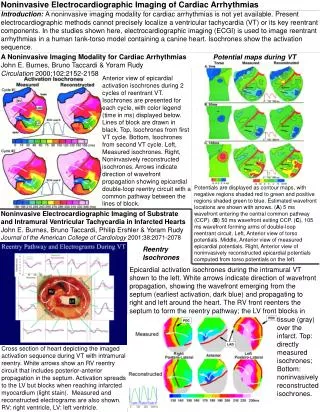
Importance of EKG Interpretation Back To Index • The underlying motivation and inspiration for this tutorial lies in the fact that the early diagnosis of heart arrhythmias will save lives. Heart Arrhythmias are at fault for the degradation of thousands of lives. Many of those affected by these arrhythmias do not even know that they have them, and therefore, are not being treated. As the treatment for heart disease continues to progress, the diagnosis of the arrhythmias becomes more and more important. By catching heart arrhythmias through EKG’s, the lives of thousands of people can be improved. Diagnosis of Cardiac Arrhythmias Through EKGs, By Greg Haider, EE5811 Biomedical Instrumentation
Back To Index Review of the Basics The Heart is composed of 4 Chambers. Each having a specific job in the cardiac cycle (click on the chart for more information) Diagnosis of Cardiac Arrhythmias Through EKGs, By Greg Haider, EE5811 Biomedical Instrumentation
Review of the Basics Back Back To Index • The Right Atrium • The Atriums are the gathering chambers of the heart • The Right Atrium performs the job of gathering oxygen depleted blood from the body. • Blood becomes oxygen depleted after nourishing the body, removing carbon dioxide from capillaries, replacing it with oxygen • From the Right Atrium, blood is passed through one of the atrioventricular valves, the tricuspid, into the right ventricle. Diagnosis of Cardiac Arrhythmias Through EKGs, By Greg Haider, EE5811 Biomedical Instrumentation
Back Back To Index Review of the Basics • The Left Atrium • The Atriums are the gathering chambers of the heart • The Left Atrium performs the job of gathering oxygen rich blood from the lungs. • Oxygen rich blood is used to nourish the body, removing carbon dioxide from capillaries, replacing it with oxygen • From the Left Atrium, blood is passed through one of the atrioventricular valves, the Mitral valve, into the left ventricle. Diagnosis of Cardiac Arrhythmias Through EKGs, By Greg Haider, EE5811 Biomedical Instrumentation
Back Back To Index Review of the Basics • The Right Ventricle • The ventricles are the pumping chambers of the heart • The Right Ventricle performs the job of pumping oxygen depleted blood to lungs • The lungs oxygenate the blood, so that it can be used again by the body for nourishing • From the Right Ventricle, blood is passed through one of the semilunar valves, the pulmonary valve, into the pulmonary artery, to the lungs Diagnosis of Cardiac Arrhythmias Through EKGs, By Greg Haider, EE5811 Biomedical Instrumentation
Back Back To Index Review of the Basics • The Left Ventricle • The ventricles are the pumping chambers of the heart • The Left Ventricle performs the job of pumping oxygenated blood to the body • Oxygenated blood is sent to the body so that it can nourish the body, removing carbon dioxide from the capillaries • From the Left Ventricle, blood is passed through one of the semilunar valves, the aortic valve, into the Aorta, to the body Diagnosis of Cardiac Arrhythmias Through EKGs, By Greg Haider, EE5811 Biomedical Instrumentation
The Motor of the Cardiovascular System Back To Index • Perimeter Layers of the Heart • Characteristics of the Myocardium • Depolarization/Repolarization • Hearts Pacemakers • Conduction Path Diagnosis of Cardiac Arrhythmias Through EKGs, By Greg Haider, EE5811 Biomedical Instrumentation
The Motor of the Cardiovascular System Back Back To Index • Perimeter Layers of the Heart • Pericardium and Epicardium • Outer 2 layers of the heart • Create a sack to hold pericardial fluid • Like oil of an engine, the liquid center creates a frictionless environment for the heart to pump, minimizing irritation • Through a fibrous connection, the epicardium attaches to the myocardium • Myocardium • Work Horse of the heart, the muscle • Endocardium • Frictionless inner layer, allowing blood to move as efficient as possible through the heart and valves Diagnosis of Cardiac Arrhythmias Through EKGs, By Greg Haider, EE5811 Biomedical Instrumentation
Back Back To Index The Motor of the Cardiovascular System • Characteristics of the Myocardium • The myocardium consists of Myocardial Cells • These cells give the myocardium the “fuel” for pumping • From these cells, the electrical stimulus in the heart begin • The cells branch and interlock such that when one cell is excited, the adjacent cell is exited, causing a chain reaction (domino effect) • The stimulated myocardial cells descend down the heart, causing the heart to contract and circulate blood Diagnosis of Cardiac Arrhythmias Through EKGs, By Greg Haider, EE5811 Biomedical Instrumentation
The Motor of the Cardiovascular System Back Back To Index • Depolarization/Repolarization • Electrically charged Ions initiate the electrical pulses in the cells • sodium ions (Na) • calcium ions (Ca) • potassium ions (K) • When at rest (Not Contracted) Myocardial cells are negatively charged, consisting of internal potassium ions and external sodium and calcium ions • Depolarization • Depolarization is the contraction of the heart, caused by fast moving Na ions and slow moving Ca ions begin to move inward, while K ions move out • Repolarization • When releasing, Na ions and Ca ions begin to trade with K ions to the normal state, this is known as repolarization • Together, Depolarization and Repolarization create the Action Potential Plot Next Diagnosis of Cardiac Arrhythmias Through EKGs, By Greg Haider, EE5811 Biomedical Instrumentation
Back Back To Index The Motor of the Cardiovascular System • Action Potential Caused by moving ions in the myocardial cells • Phase 0: • Rapid depolarization of Na ions • Phase 1 and 2: • Slow depolarization of Ca ions, start of repolarization of K ions • Phase 3: • Complete repolarization • Phase 4: • At rest Diagnosis of Cardiac Arrhythmias Through EKGs, By Greg Haider, EE5811 Biomedical Instrumentation
Back Back To Index The Motor of the Cardiovascular System • Pacemakers • nodes of myocardial cells that do not rest, but continue to depolarize and repolarize at an inherent rate starting the conduction through the heart • Sinoatrial Node (SA) – main pace maker • Automaticiy Foci – Backup pacemakers, pace if a higher pacemaker fails to beat • Atrial foci: 60 – 80 bpm • Junctional foci: 40 – 60 bpm • Ventricular foci: 20 – 40 bpm Diagnosis of Cardiac Arrhythmias Through EKGs, By Greg Haider, EE5811 Biomedical Instrumentation
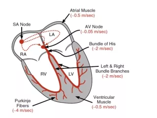
Back Back To Index Conduction Path Click on the illustration for description Purkinje Fibers Diagnosis of Cardiac Arrhythmias Through EKGs, By Greg Haider, EE5811 Biomedical Instrumentation
Back Back To Index Conduction Path • Start of conduction path • SA node is located in the upper right atrium • This is the main pacemaker of heart, kick starting the depolarization of the atriums, forcing blood into the ventricles Purkinje Fibers Diagnosis of Cardiac Arrhythmias Through EKGs, By Greg Haider, EE5811 Biomedical Instrumentation
Back Back To Index Conduction Path • Atrioventricular Node • The AV node produces a pause in the electrical flow through the heart, allowing the atria to completely contract and fill the ventricles Purkinje Fibers Diagnosis of Cardiac Arrhythmias Through EKGs, By Greg Haider, EE5811 Biomedical Instrumentation
Back Back To Index Conduction Path • His Bundle • The His Bundle conducts the depolarization down the septum into the ventricles and branches off into the right and left Bundle Branches Purkinje Fibers Diagnosis of Cardiac Arrhythmias Through EKGs, By Greg Haider, EE5811 Biomedical Instrumentation
Back Back To Index Conduction Path • Purkinje Fibers • These fibers stem off of the right and left bundle branches into the myocardium producing a strong explosion of current, pumping blood out of the heart Purkinje Fibers Diagnosis of Cardiac Arrhythmias Through EKGs, By Greg Haider, EE5811 Biomedical Instrumentation
Back To Index EKG Measurement Process • The 12 lead EKG is the Most common form of EKG Measurement • This form of measurement consist so 6 limp leads 6 chest leads • The 6 limb leads are created with electrodes attached to both arms and the left foot • 3 Bipolar Limb leads • Each lead acts as both anode and cathode • 3 Augmented Limb leads • Two leads set as anode, one as the cathode • 6 chest leads • Positioned along the chest surrounding the heart • All chest leads are positively charged, allowing detection of the hearts electrical activity Next Diagnosis of Cardiac Arrhythmias Through EKGs, By Greg Haider, EE5811 Biomedical Instrumentation
Back Back To Index EKG Measurement Process • The 12-lead ECG allows the depolarization and depolarization of the heart to be detected • Depolarization of the Atrium produces the P-Wave on the EKG printout • The complex QRS-Wave of the printout is produced through ventricular depolarization • Finally, the EKG’s T-Wave is produced when the ventricular repolarizes. Diagnosis of Cardiac Arrhythmias Through EKGs, By Greg Haider, EE5811 Biomedical Instrumentation
Back To Index Causes of Heart Arrhythmias • Unknown causes • Too intermittent to diagnose • Weak or lack of connection between Myocardial cells • Caused by dead tissues or scars, infarctions • Usually caused by extreme electrical impulses such as a heart attack, also known as Myocardial Infarction • Chemical Imbalances • Because the depolarization and repolarization of the heart relies the movement of ions, and imbalance of these ions can produce abnormal beats • Sodium imbalance • Calcium imbalance • Electrical Confusion • Again, usually caused by extreme electrical impulses that unsynchronize the system, sometimes to the point that it is unrecoverable Diagnosis of Cardiac Arrhythmias Through EKGs, By Greg Haider, EE5811 Biomedical Instrumentation
Back To Index Diagnosis of Arrhythmias Using the EKG • Arrhythmias • First Degree Heart Block • Second Degree Heart Block • Sinus Rhythm Bradycardia and Tachycardia • Hypercalcemia • Hyperkalemia • Idioventricular Rhythm • Premature Atrial Contraction • Premature Ventricular Contraction • Ventricular Tachycardia • Ventricular Fibrillation • Asystole Diagnosis of Cardiac Arrhythmias Through EKGs, By Greg Haider, EE5811 Biomedical Instrumentation
Back Back To Index Diagnosis of Arrhythmias Using the EKG • First Degree Heart Block Normal EKG First Degree Heart Block • First-degree heart block, or first-degree AV block, is the condition under which the electrical impulse moves through the AV node at a slower rate than normal. The cause of first degree heart block is not always known since in some cases it can be intermittent. However, in many cases, it may be to a weakened conduction path caused by minor scarring of the tissue. • The time it takes for the impulse to get from the atria to the ventricles (the PR interval) should be less than about 0.2 seconds for a normal rhythm. If this time is longer on the EKG, than the patient under test is experiencing first-degree heart block Diagnosis of Cardiac Arrhythmias Through EKGs, By Greg Haider, EE5811 Biomedical Instrumentation
Back Back To Index Diagnosis of Arrhythmias Using the EKG • Second Degree Heart Block Normal EKG Second Degree Heart Block • While first degree heart block may cause a physician to take a second look, it is usually not harmful, producing a valid rate. However, second-degree heart block paints a different picture. In second-degree heart block, the AV junction moves so slow that some of the electrical impulses from the atrium never reach the ventricles, meaning scarring is bad enough to completely disrupt the electrical connection. This behavior will cause the EKG to produce a double P-wave. Diagnosis of Cardiac Arrhythmias Through EKGs, By Greg Haider, EE5811 Biomedical Instrumentation
Back Back To Index Diagnosis of Arrhythmias Using the EKG • Sinus Rhythm Bradycardia and Tachycardia Normal EKG Bradycardia Tachycardia • Sinus Rhythm Bradycardia is the condition in which the patient’s rhythm is under 60 beats per minute. This condition is usually caused by the SA node firing too slow but could also be a sign that another atrial foci has taken over pacing due to scarred tissue. However, the blockage of scar tissue can usually be determined by the characteristics of the P wave, and would not be classified under this condition. During sinus rhythm bradycardia, the R to R interval is lengthened, and at times, the P wave is widened. • Sinus Rhythm Tachycardia is the opposite of Bradycardia, in which the internal heart rate is faster than that of a normal rate. During this condition, the SA node is firing too fast. When a patient is experiencing Sinus Tachycardia the heart rate of that patient is greater than 100bpm. Diagnosis of Cardiac Arrhythmias Through EKGs, By Greg Haider, EE5811 Biomedical Instrumentation
Back Back To Index Diagnosis of Arrhythmias Using the EKG • Hypercalcemia Normal EKG Hypercalcemia • Hypercalcemia is a condition under which a patient is experiencing high levels of calcium in his/her body, a chemical imbalance. Because calcium plays such an important role in the behavior of the heart, excess calcium can cause it to perform abnormally. With too much calcium, the interval between the final depolarization phase caused by calcium ions and the repolarization phase is shortened. The effects of Hypercalcemia are very difficult to see in some patients, however with a high enough content, the calcium can affect the heart such that the S-T interval is visibly shortened, or even invisible. Diagnosis of Cardiac Arrhythmias Through EKGs, By Greg Haider, EE5811 Biomedical Instrumentation
Back Back To Index Diagnosis of Arrhythmias Using the EKG • Hyperkalemia Normal EKG Hyperkalemia • Hyperkalemia is the excessive amount of potassium in the body. Again, like Calcium, Potassium is an important ion in the heart, affecting the behaviors of the hearts depolarization. Therefore, large amounts of potassium in a patient’s body will affect the EKG. When large amounts of Potassium are found in the body, the T-wave takes on a tent shaped form. As with Hypercalcemia, Hyperkalemia is a very difficult condition to diagnose using an EKG printout due to the small effects that are produced with the additional Ions. Diagnosis of Cardiac Arrhythmias Through EKGs, By Greg Haider, EE5811 Biomedical Instrumentation
Back Back To Index Diagnosis of Arrhythmias Using the EKG • Idioventricular Rhythm Normal EKG Idioventricular Rhythm • When one the ectopic foci, located at or above the atrioventricular node, fails to discharge, usually due to infracted tissue, atrial contraction never occurs. When one of these foci fails to fire, a foci node within the ventricle will takeover, producing ventricular depolarization and repolarization. With the loss of Atrial contraction, the P-wave is absent from the EKG, showing only the QRS complex. Also, because the pacemaker is located in the ventricle, the QRS wave takes on a much different rate than that seen on a normal EKG. Because a separate automaticity foci is controlling the rate, the rates of Idioventricular Rhythms are very slow – usually 30 to 40 beats per minute. Diagnosis of Cardiac Arrhythmias Through EKGs, By Greg Haider, EE5811 Biomedical Instrumentation
Back Back To Index Diagnosis of Arrhythmias Using the EKG • Premature Atrial Contraction Normal EKG Premature Atrial Contraction • In many cases, physicians are unable to determine the cause of Premature Atrial Contraction (PAC), however the signs of this condition are obvious on the EKG. In PAC, an SA node kicks off atrial contraction soon after ventricular repolarization, producing a strange T-P interval from one beat to the next. The heart rate usually picks up as normal, but with a shift in phase. Diagnosis of Cardiac Arrhythmias Through EKGs, By Greg Haider, EE5811 Biomedical Instrumentation
Back Back To Index Diagnosis of Arrhythmias Using the EKG • Premature Ventricular Contraction Normal EKG Premature Ventricular Contraction • Premature Ventricular Contraction is the escape of a ventricular automaticity foci pace, while the SA or other ectopic foci are performing their duties correctly; there is no block that makes it necessary for this foci to fire. In this case, the ventricle contracts before the atrial pacemaker has an opportunity to initiate the normal rate. Therefore, this condition is seen on the EKG as a missed P-wave, and an irregular, widened QRS complex. Diagnosis of Cardiac Arrhythmias Through EKGs, By Greg Haider, EE5811 Biomedical Instrumentation
Back Back To Index Diagnosis of Arrhythmias Using the EKG • Ventricular Tachycardia Normal EKG Ventricular Tachycardia • Ventricular tachycardia is defined as three or more beats of ventricular origin in succession at a rate greater than 100 beats per minute. There are no normal-looking QRS complexes and the rhythm is usually regular, but on occasion it may be modestly irregular. Much like PVC, under the condition of ventricular tachycardia, the SA is still depolarizing the atria, however in ventricular tachycardia a ventricular foci node is controlling the pace at a rate faster or equal to that of the atria. Because the ventricle is in refractory when the atria depolarization is passed to the atrioventricular node, this stimulus has no effect on the synching the two rates. The ventricular depolarization far outweighs the atria depolarization, dominating the EKG screen. Diagnosis of Cardiac Arrhythmias Through EKGs, By Greg Haider, EE5811 Biomedical Instrumentation
Back Back To Index Diagnosis of Arrhythmias Using the EKG • Ventricular Fibrillation Normal EKG Ventricular Fibrillation • Ventricular fibrillation is caused by the total disorder of ventricular foci. This condition is caused by extreme electrical stress in the heart, scrambling the rhythm of the heart. Under these conditions, the ventricle is attempting to contract at different rates, at different locations. This chaotic depolarization results in a ventricular fibrillation, or a quiver, rather than a rhythm, resulting in very little blood flow from the heart. Ventricular fibrillation is shown on the EKG as very small, random pulses. Diagnosis of Cardiac Arrhythmias Through EKGs, By Greg Haider, EE5811 Biomedical Instrumentation
Back Back To Index Diagnosis of Arrhythmias Using the EKG • Asystole Normal EKG Asystole • The simplest EKG reading to determine is that of Asystole, or death. At the time of Asystole, the heart is fully depolarized, in both the atrial and the ventricular, with no attempts to repolarize. Because there is no electrical activity in the heart, there is no EKG surge caused by the lead connections. The EKG reading is just what one would expect under these conditions, flat. Diagnosis of Cardiac Arrhythmias Through EKGs, By Greg Haider, EE5811 Biomedical Instrumentation
Back To Index Medical Ethics of EKG Diagnosis • The EKG process itself appears harmless, however every procedure involves an ethical decision • Ethical Question: • Is it necessary? • May Cause Physical Discomfort • Leads must be attached to the body • May Cause Mental Discomfort • Embarrassment • Fear Next Diagnosis of Cardiac Arrhythmias Through EKGs, By Greg Haider, EE5811 Biomedical Instrumentation
Back Back To Index Medical Ethics of EKG Diagnosis • Ethics of treating arrhythmias following a Diagnosis • Comfort • Drug treatment versus possible implantable device • Life Circumstances • Will this require monthly checkups? • Single parent • Traveling position • Fear of hospitals • Age • Elderly • Would surgery produce more risk than the condition itself? • Young • Because patient is still growing, should other alternative treatment be decided on if an implantable is the usual treatment? • Finances • Is the patient insured? • Will the patient be retiring, will they keep insurance? • Many heart conditions are chronic, therefore the treatment will be with them forever, can they not only afford it now, but what about 10, 20, or even 50 years down the road • Each decision must be made on a patient to patient bases • The best treatment is not always the right treatment • Do good versus Do right Diagnosis of Cardiac Arrhythmias Through EKGs, By Greg Haider, EE5811 Biomedical Instrumentation