Download
1 / 1
10 likes | 96 Views
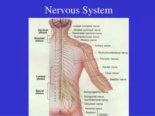
Periodic Leg Movements in Spinal Cord Injury: Evaluation of Arousals and Treatment Effect Aaro Salminen 1 , Mauro Manconi 2 , Ville Rimpilä 1 , Teemu Luoto 3 , Raffaele Ferri 4 , Olli Polo 1,3 1) University of Tampere, Finland 2) Civic Hospital of Lugano, Switzerland

E N D
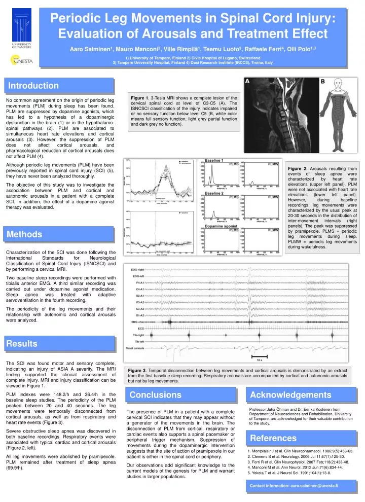
Periodic Leg Movements in Spinal Cord Injury: Evaluation of Arousals and Treatment Effect Aaro Salminen1, Mauro Manconi2, Ville Rimpilä1, Teemu Luoto3, Raffaele Ferri4, Olli Polo1,3 1) University of Tampere, Finland 2) Civic Hospital of Lugano, Switzerland 3) Tampere University Hospital, Finland 4) Oasi Research Institute (IRCCS), Troina, Italy Introduction Figure 1. 3-Tesla MRI shows a complete lesion of the cervical spinal cord at level of C3-C5 (A). The ISNCSCI classification of the injury indicates impaired or no sensory function below level C5 (B, white color means full sensory function, light grey partial function and dark grey no function). No common agreement on the origin of periodic leg movements (PLM) during sleep has been found. PLM are suppressed by dopamine agonists, which has led to a hypothesis of a dopaminergic dysfunction in the brain (1) or in the hypothalamo-spinal pathways (2). PLM are associated to simultaneous heart rate elevations and cortical arousals (3). However, the suppression of PLM does not affect cortical arousals, and pharmacological reduction of cortical arousals does not affect PLM (4). Although periodic leg movements (PLM) have been previously reported in spinal cord injury (SCI) (5), they have never been analyzed thoroughly. The objective of this study was to investigate the association between PLM and cortical and autonomic arousals in a patient with a complete SCI. In addition, the effect of a dopamine agonist therapy was evaluated. Figure 2. Arousals resulting from events of sleep apnea were characterized by heart rate elevations (upper left panel). PLM were not associated with heart rate elevations (lower left panel). However, during baseline recordings, leg movements were characterized by the usual peak at 20-30 seconds in the distribution of inter-movement intervals (right panels). The peak was suppressed by pramipexole. PLMS = periodic leg movements during sleep, PLMW = periodic leg movements during wakefulness. Methods Characterization of the SCI was done following the International Standards for Neurological Classification of Spinal Cord Injury (ISNCSCI) and by performing a cervical MRI. Two baseline sleep recordings were performed with tibialis anterior EMG. A third similar recording was carried out under dopamine agonist medication. Sleep apnea was treated with adaptive servoventilation in the fourth recording. The periodicity of the leg movements and their relationship with autonomic and cortical arousals were analyzed. Results The SCI was found motor and sensory complete, indicating an injury of ASIA A severity. The MRI finding supported the clinical assessment of complete injury. MRI and injury classification can be viewed in Figure 1. PLM indexes were 148.2/h and 36.4/h in the baseline sleep studies. The periodicity of the PLM peaked between 20 and 40 seconds. The leg movements were temporally disconnected from cortical arousals, as well as from respiratory and heart rate events (Figure 3). Severe obstructive sleep apnea was discovered in both baseline recordings. Respiratory events were associated with typical cardiac and cortical arousals (Figure 2, left). All leg movements were abolished by pramipexole. PLM remained after treatment of sleep apnea (69.9/h). Figure 3. Temporal disconnection between leg movements and cortical arousals is demonstrated by an extract from the first baseline sleep recording. Respiratory arousals are accompanied by cortical and autonomic arousals but not by leg movements. Conclusions Acknowledgements Professor Juha Öhman and Dr. Eerika Koskinen from Department of Neurosciences and Rehabilitation, University of Tampere, are acknowledged for their valuable contribution to the study. The presence of PLM in a patient with a complete cervical SCI indicates that they may appear without a generator of the movements in the brain. The disconnection of PLM from cortical, respiratory or cardiac events also supports a spinal pacemaker or peripheral trigger mechanism. Suppression of movements during the dopaminergic intervention suggests that the site of action of pramipexole in our patient is either in the spinal cord or periphery. Our observations add significant knowledge to the current models of the genesis for PLM and warrant studies in larger populations. References 1. Montplaisir J et al. Clin Neuropharmacol. 1986;9(5):456-63. 2. Clemens S et al. Neurology. 2006 Jul 11;67(1):125-30. 3. Ferri R et al. Clin Neurophysiol. 2007 Feb;118(2):438-48. 4. Manconi M et al. Ann Neurol. 2012 Jun;71(6):834-44. 5. Yokota T et al. J Neurol Sci. 1991;104(1):13-8. Contact information: aaro.salminen@unesta.fi