Download
1 / 31
530 likes | 2k Views
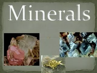
Minerals. According to the body needs, minerals are divided into 2 groups:. I. Macrominerals:. They are required in amounts greater than 100 mg/day. They include: calcium, phosphorus, magnesium, sodium, potassium and chloride. II. Microminerals (trace elements):.

E N D
Minerals According to the body needs, minerals are divided into 2 groups: I. Macrominerals: They are required in amounts greater than 100 mg/day. They include: calcium, phosphorus, magnesium, sodium, potassium and chloride II. Microminerals (trace elements): They are required in amounts less than 100 mg/day. They include: iron, copper, fluoride, iodine, manganese, selenium and zinc.
Macrominerals Calcium Sources: Milk, milk products, beans and egg yolk. Absorption: By active transport system in the upper small intestine by the help of vitamin D (1,25 dihydroxy cholicalciferol). Body calcium: calcium is the most abundant mineral in the body. Most of calcium (99%) is present in the skeleton (bones and teeth), the remaining 1% are present in body fluids and other tissues Plasma calcium: normal level ranges from 9-11 mg/dl
Hormonal regulation of Plasma calcium: 1. Calcitonin: is secreted from thyroid gland in response to increased blood Ca level. It decreases Ca through: • Mobilization of calcium from blood into bone • Decrease Ca reabsorption by renal tubules 2. Parathyroid hormone (PTH): secreted from parathyroid gland in response to decreased calcium levels. It increase calcium by: • Mobilization of calcium from bone to blood (bone resorption) • Increases Ca reabsorption by renal tubules • Increase absorption of calcium from small intestine through stimulation of vitamin D activation.
Factors affecting calcium absorption • A- Factors affecting Ca absorption: 1-High protein diet: amino acid forms soluble salt with calcium. 2-Ph: an acidic PH is essential for calcium absorption. 3- High lactate or citrate that form soluble salts with calcium.
Factors affecting calcium absorption B- Factors inhibiting Ca absorption: 1- High diet phosphate, oxalate, phytate form insoluble salt with CA. 2- Alkalinity: Ca absorption( treatment of peptic ulcer). 3- Fatty acid: insoluble Ca soaps with Ca.
Function of Calcium • 1- Unionized Calcium: A- It enters in the structure of bone and teeth. 2- Ionized calcium: A- Transmission of nerve impulses. B-Contraction of muscles. C- Decrease of neuromuscular excitability. Deficiency of ionized calcium lead to tetany. D- Blood and milk clotting. E-Maintenance of cell membrane permeability. F- Activation of certain enzyme e.g. pyruvate kinase. G- Medication of some hormone responses e.g. it acts together with calmodulin as third messenger for hormone depending on cyclic AMP.
Hypercacemia: may be due to: • Hyperparethyrodism due to adenoma ( benign tumor of the gland) • Excess intake of vitamin D or calcium • Milk-alkali syndrome : patients who receive milk and alkalies for long time, for treatment of peptic ulcer • Drugs such as thiazide diuretics Hypocacemia: due to: • Hypoparathyrodism • Renal disease where activation of vitamin D is inhibited Requirments: • Adult: 800 mg/day • Children, pregnant and lactating women: 800-1200 mg/day
Phosphorus • Source: Milk meat and leafy vegetables • Absorption:By active transorport and regulated by Vitamin D • Factors affect of ca absorption also affect that of Phosphorus. • Body Phosphorus : Most of it present in bone and teeth. In the form hydroxy apatite: 3 Ca3(PO4)2.Ca(OH)2 • Blood phosphorus 3-5 mg/dl.
Factors Affecting blood phosphorus • 1- Parathyroid hormone: A- PTH decrease blood phosphorus by stimulating its excretion (through inhibiting its renal tubular re absorption). • 2- Active Vitamin D (calcitriol): • A- Hypophosphatemia stimulates directly the renal hydroxylation of 25 (OH)D3 into 1,25 (OH)D3 (Calcitriol). • B- Calcitriol increases blood phosphorus through stimulation of: • 1- Absorption of phosphorus from the intestine . • Bone resorption i.e. mobilization of phosphorus from bone. • Renal reabsorption by renal tubules.
Function of Phosporus 1-It enters in the structure of bone and teeth. 2- It enters in the structure of the following cellular components: a- Nucleic acids DNA RNA B- Phospholipids e.g. lecithin , cephalin. C- Phosphproteins. D- Coenzyme e.g. NAD, NADP. E- High energy phospate compounds e.g Atp, GTP,creatine phosphate. F- Cyclic Amp and Cuclic GTP. G- Carbohydrate intermediates e.g. glucose 6 phospate, fructose -1-p. 3- it enters in the formation of blood buffers.
Sodium Sources: The main source is table salt Absorption: from small intestine (ileum). It is nearly completely absorbed. Body sodium: 2/3 of sodium is present in tissues and body fluids. About 1/3 is present in skeleton (bone and teeth) Requirements: 5 g/day Alterations of plasma sodium: Hypernatremia: excess plasma sodium is caused by: • Cushing syndrome. • Conn's disease due to excessive aldosterone secretion • Diabetes inspidus due to rapid loss of water • Drugs such as cortisone
Hyponatremia: decrease plasma sodium caused by: • Addison's disease: due to deficiency of aldosterone • Renal failure where renal reabsorption of sodium is inhibited • Dehydration: due to loss of water and sodium • Thaizide diuretics which block renal reabsorption of sodium Toxicity of sodium: Hypertension in susceptible individuals. Functions: 1- maintentance of osmotic pressure and volume of plasma and extracellular fluid. 2-Transmission of nerve impulses. 3-Contraction of muscle. 4-Regulation of acid base balance.
Potassium • Sources: • vegetables, fruits and nuts • Absorption: • Readily occur from small intestine • Body K: • 2/3 of potassium is present in tissues and body fluids. About 1/3 is present in skeleton (bone and teeth) • Requirements: 4 g/day Functions of potassium: The same of sodium except it act in intercellular fluid.
Alterations of plasma potassium: Hyperkalemia: excess plasma potassium is caused by: • Addison's disease: due to deficiency of aldosterone • Acidosis • Tissue necrosis e.g. major trauma and burns due to leakage of tissue potassium • Chronic renal failure with oliguria • Uncontrolled D. M. Lack of insulin prevent potassium from entering cells Hypokalemia: decrease plasma potassium caused by: • alkalosis • treatment of hyperglycemia with insulin without taking potassium, as insulin helps potassium ion to enter cells • Excessive vomiting and diarrhea • Cushing syndrome • Diuretics therapy.
Chloride Functions: • Chloride is the main extracellular anion. Together with sodium, it maintains the osmotic pressure and volume of plasma and extracellular fluid. • Chloride ions is essential for formation of HCl in the stomach. • Activation of enzyme:Cl activates salivary and pancreatic amylase enzymes.
Maganisum • Functions: 1- it enters in the structure of skeleton (bone and teeth). 2-It activates many enzyme e.g. kinase enzyme. 3- it is required for transport of other cation (Ca. Na, K) across the cell membrane . 4- it is important for muscle contraction, nerve impulse transmission and it decrease neuromuscular excitability.
Microminerals (Trace elements) IRON Sources: Liver, heart, kidney, spleen, and fish Sugarcane syrup (molasses), dates and egg yolk N.B. Most of dietary iron is present in the ferric state. Absorption: from small intestine. Usually 10-20% of dietary iron is only absorbed. Iron is absorbed in ferrous state. Reducing substances such as vitamin C and SH- of cysteine of dietary protein help the reducing of ferric ions into the absorbable form (ferrous state). Body iron: The total body iron of adult is 3-5 g distributed as follows: I -RBCs iron ( haemoglobin) is about 65% of total iron II- Tissue iron (32%): includes
Available forms (28%) i.e. can be used when there body need 1-Ferritin : is the main storage form of iron. Composed of protein (apoferritin + iron). Present in iron stores: liver, spleen, bone marrow and intestine. 2- Haemosiderin: These are granules composed of iron, protein and polysaccharides. Used as another store of iron Non available forms (4%): can not be used even there is body need. All these forms are hemoprotein i.e. contain heme ring. Examples are: • Myoglobin: present in muscle and heart • Cytochromes: a, b and c: act as electron carriers • Catalase and peroxidase: act to destroy H2O2 III- Plasma iron: present in the form of: 1-plasma iron : ranges from 60- 160 μg/ dl 2- Transferrin : is a glycoprotein which carry iron in ferric state.
Transport and storage of iron: • Absorbed iron enters in the portal blood in the ferrous state. • In the plama it is rapidly oxidized to ferric state by the help of protein- containing copper called: Ceruloplasmin • Then ferric ions are carried by transferring, which is\taken mostly by bone marrow to synthesize hemoglobin • Iron from iron stores (ferritin) can be released into plasma and carried by transferring to be used by B.M. and other tissues Requirements: • adults: 10 mg/day • Pregnant and lactating female: 30 mg/day
Copper Sources : Most diet provides the amount of copper needed per day. Absorption : Mainly in the upper small intestine Body copper : 64% of copper are found in muscles and the remaining in other tissues including liver and bones. Plasma copper : 90 μg/ dl. Functions: 1- copper is essential for: A-Homoglobuin synthesis. B- Bone formation. C- mantence of mylin of the nerve. 2-Copper is essential component of several metaloenzymes such as: A-Ceruloplasmin: which oxidizes ferrous into ferric in plasma B-Superoxide desmutase: antioxidant enzyme C-Cytochromeoxidase
3- copper activates many enzyme e.g. uricase and dopamine hydroxylase. Requirements:2-3 mg/day Alterations of plasma copper: Hyper cupremia (Excess copper and ceruloplasmin) occur in infections and malignancies Hypocupremia (decreased plasma copper):occure in a disease called Wilson's disease in which copper accumulate in large amounts in: • Liver causing liver cirrhosis • cornea causing greenish- brown color of the corneal margine Kidney causing damage of renal tubules leading to increased excretion of copper and ceruloplasmin resulting in low plasma copper.
Iodine • The only function is formation of T3 and T4 from thyroid gland, so deficiency of iodine leading to hypothyroidism and disease called Simple Goiter. • Requirements: 100-150 μg/day
Zinc Functions of zinc: • Zinc is essential for growth and reproduction. • It plays a role in tissue repair and wound healing • Zinc forms a complex with insulin in β cells of the pancreases. this helps crystallization ,storage and release of insulin. • Zinc is essential component of a number of enzymes e.g.: • A- alkaline phoshatase. • B- Carbonic anhyrase. • C- Superoxide dismutase. • D- Carboxy peptidase.
Zinc 5- Zinc is required for mobilization of vitamin A from the liver and subsequently maintain the normal concentration of vitamin A in plasma. Zinc deficiency: it causes: • Hypogonadism. • Poor healing of wounds. • Poor appetite and retard growth in children. • Liver cirrhosis.
Selenium • A_selenium is an essential component of the enzyme glutathione peroxide (GSH-PX) which catalyzes the reaction: 2 GSH+H2O2 GSH-PX GSSG +2 H2O • B- This reactions as protective mechanism agonist the oxidative damage of hydrogen peroxide (H2O2) and fatty acid hydroperoxide by destroying them: • In RBCs, it protect heamoglobin and red cell membrane. • In the liver, it is important for detoxifying liquid hydroperoxides. • In the lens tissue of the eye< it prevents its oxidative damage.
Selenium • Deficiency of selenium (GSH-PX): it causes: • Hemolytic anemia. • Liver cirrhosis • Cataract.
Manganese • A- Manganese is essential for: • Normal bone structure. • Reproduction (spermatogenesis and ovulation). • Normal function of the CNS. • B- Manganese is component of: • Pyruvatecarboxylase enzyme. • Superoxide dismutase enzyme. • C- Manganese activates the arginase enzyme.
Cobalt • A- coblet is a component of Vitamin B12 which is nessary for normal blood cell formation. • B- Cobalt gives vitamin B12 its red colour. • C- Enzyme requrining Vitamin B12 for their activites are: • Methylmalonyl COA mutase. • Methyletrahydrofolateoxidoreductase. • Homocysteninemethytransferase. • Ribonuclotidereductase. • D- Deficiency of Vitamin B12 causes pernicious anaemia .
Chromium • It acts only together with insulin to promote glucose utilization. • Its deficiency leads to impairment of glucose utilization by tissue.
Molybdenum • It is a component of oxidase enzymes e.g.Xanthine oxidase.
Flouride • It increase the hardness of bone and teeth. • Its deficiency causes dental carries and osteoporosis. • Nowadays, it is supplied in drinking water. • Excess fluoride leads to fluorosis: mottling and discoloration of the enamel of teeth and changes in bones.