Download
1 / 53
530 likes | 685 Views
Human Reproductive System Part I. Chapter 16 BIO 160 Kelly Trainor. The Reproductive System. Gonads—primary sex organs Testes in males Ovaries in females Gonads produce gametes (sex cells) and secrete hormones Sperm—male gametes Ova (eggs)—female gametes. Male Reproductive System.

E N D
Human Reproductive SystemPart I Chapter 16 BIO 160 Kelly Trainor
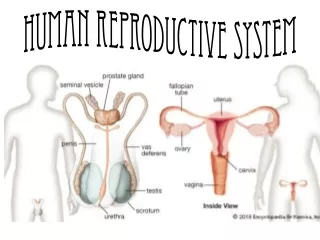
The Reproductive System • Gonads—primary sex organs • Testes in males • Ovaries in females • Gonads produce gametes (sex cells) and secrete hormones • Sperm—male gametes • Ova (eggs)—female gametes
Male Reproductive System • Testes • Duct system • Epididymis • Ductus (vas) deferens • Urethra • Accessory organs • Seminal vesicles • Prostate • Bulbourethral glands • External genitalia • Penis • Scrotum
Male Reproductive System Figure 16.2a
Male Reproductive System Figure 16.2b
Testes Figure 16.1
Testes • Each lobule contains one to four seminiferous tubules • Tightly coiled structures • Function as sperm-forming factories • Interstitial cells in the seminiferous tubules produce androgens such as testosterone
Epididymis • Comma-shaped, tightly coiled tube • Found on the superior part of the testis and along the posterior lateral side • Functions to mature and store sperm cells (at least 20 days) • Expels sperm with the contraction of muscles in the epididymis walls to the vas deferens
Ductus Deferens (Vas Deferens) • Carries sperm from the epididymis to the ejaculatory duct • Passes through the inguinal canal and over the bladder • Moves sperm by peristalsis • Spermatic cord—ductus deferens, blood vessels, and nerves in a connective tissue sheath • Ends in the ejaculatory duct which unites with the urethra • Ejaculation—smooth muscle in the walls of the ductus deferens create peristaltic waves to squeeze sperm forward • Vasectomy—cutting of the ductus deferens at the level of the testes to prevent transportation of sperm
Urethra • Extends from the base of the urinary bladder to the tip of the penis • Carries both urine and sperm • Sperm enters from the ejaculatory duct
Accessory Organs • Seminal vesicles • Prostate • Bulbourethral glands
Seminal Vesicles • Located at the base of the bladder • Produces a thick, yellowish secretion (60% of semen) • Fructose (sugar) • Vitamin C • Prostaglandins • Other substances that nourish and activate sperm Prostate • Encircles the upper part of the urethra • Secretes a milky fluid • Helps to activate sperm • Enters the urethra through several small ducts
Bulbourethral Glands • Pea-sized gland inferior to the prostate • Produces a thick, clear mucus • Cleanses the urethra of acidic urine • Serves as a lubricant during sexual intercourse • Secreted into the penile urethra
Prostate Figure 16.2a
Semen • Mixture of sperm and accessory gland secretions • Advantages of accessory gland secretions • Fructose provides energy for sperm cells • Alkalinity of semen helps neutralize the acidic environment of vagina • Semen inhibits bacterial multiplication • Elements of semen enhance sperm motility
External Genitalia • Scrotum • Divided sac of skin outside the abdomen • Maintains testes at 3°C lower than normal body temperature to protect sperm viability • Penis • Delivers sperm into the female reproductive tract • Regions of the penis • Shaft • Glans penis (enlarged tip) • Prepuce (foreskin) • Folded cuff of skin around end • Often removed by circumcision • Internally there are three areas of spongy erectile tissue around the urethra • Erections occur when this erectile tissue fills with blood during sexual excitement
External Genitalia Figure 16.2a
Spermatogenesis • Production of sperm cells • Begins at puberty and continues throughout life • Occurs in the seminiferoustubules • Spermatogonia (stem cells) undergo rapid mitosis to produce more stem cells before puberty • Follicle-stimulating hormone (FSH) modifies spermatogonia division • Primary spermatocytes undergo meiosis • One primary spermatocyte produces four haploid spermatids • Spermatids—23 chromosomes (half as much material as other body cells)
Human Life Cycle • Union of a sperm (23 chromosomes) with an egg (23 chromosomes) creates a zygote (2n or 46 chromosomes)
Spermiogenesis • Late spermatids are produced with distinct regions • Head • Midpiece • Tail • Sperm cells result after maturing of spermatids • Spermatogenesis (entire process, including spermiogenesis) takes 64 to 72 days
Anatomy of a Mature Sperm Cell • The only human flagellated cell • Head • Contains DNA • Acrosome—“helmet” on the nucleus, similar to a large lysosome • Breaks down and releases enzymes to help the sperm penetrate an egg • Midpiece • Wrapped by mitochondria for ATP generation
Testosterone Production • The most important hormone of the testes • Functions of testosterone • Stimulates reproductive organ development • Underlies sex drive • Causes secondary sex characteristics • Deepening of voice • Increased hair growth • Enlargement of skeletal muscles • Thickening of bones
Human Reproductive SystemPart II Chapter 16 BIO 160 Kelly Trainor
Female Reproductive System • Ovaries • Duct System • Uterine tubes (fallopian tubes) • Uterus • Vagina • External genitalia
Female Reproductive System Figure 16.8b
Ovaries • Composed of ovarian follicles (sac-like structures) • Each follicle consists of • Oocyte (immature egg) • Follicular cells—surround the oocyte
Ovarian Follicle Stages • Primary follicle—contains an immature oocyte • Graafian (vesicular) follicle—growing follicle with a maturing oocyte • Ovulation—when the egg is mature, the follicle ruptures; occurs about every 28 days • The ruptured follicle is transformed into a corpus luteum
Uterine (Fallopian) Tubes • Receive the ovulated oocyte • Provide a site for fertilization • Attach to the uterus • Little or no contact between ovaries and uterine tubes • Supported and enclosed by the broad ligament
Uterine Tube Anatomy and Physiology • Fimbriae • Finger-like projections at the distal end of the uterine tube • Receive the oocyte from the ovary • Cilia • Located inside the uterine tube • Slowly move the oocyte towards the uterus (takes 3–4 days) • Fertilization occurs inside the uterine tube since oocyte lives about 24 hours
Female Reproductive System Figure 16.8b
Uterus • Located between the urinary bladder and rectum • Hollow organ • Regions of the uterus • Body—main portion • Fundus—superior rounded region above where uterine tube enters • Cervix—narrow outlet that protrudes into the vagina • Functions of the uterus • Receives a fertilized egg • Retains the fertilized egg • Nourishes the fertilized egg
Female Reproductive System Figure 16.8b
Walls of the Uterus • Endometrium • Inner layer • Allows for implantation of a fertilized egg • Sloughs off if no pregnancy occurs (menses) • Myometrium—middle layer of smooth muscle • Perimetrium (visceral peritoneum)—outermost serous layer of the uterus
Female Reproductive System Figure 16.8b
Vagina • Extends from cervix to exterior of body • Located between bladder and rectum • Serves as the birth canal • Receives the penis during sexual intercourse
External Genitalia • Mons Pubis • Fatty area overlying the pubic symphysis • Covered with pubic hair after puberty • Labia—skin folds • Labia majora—hair-covered skin folds • Labia minora—delicate, hair-free folds of skin • Clitoris • Contains erectile tissue • Corresponds to the male penis • The clitoris is similar to the penis in that it is • Hooded by a prepuce • Composed of sensitive erectile tissue • Becomes swollen with blood during sexual excitement
Vestibule and Greater Vestibular Glands • Vestibule • Enclosed by labia majora • Contains external openings of the urethra, vagina • Greater vestibular glands • One is found on each side of the vagina • Secretes lubricant during intercourse
External Genitalia (Vulva) Figure 16.9
Human Reproductive SystemPart III Chapter 16 BIO 160 Kelly Trainor
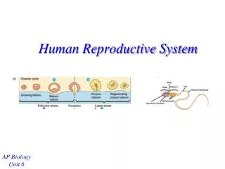
Oogenesis and the Ovarian Cycle • The total supply of eggs are present at birth • Ability to release eggs begins at puberty • Reproductive ability ends at menopause • Oocytes are matured in developing ovarian follicles • Oogonia—female stem cells found in a developing fetus • Oogonia undergo mitosis to produce primary oocytes
Oogenesis and the Ovarian Cycle • Primary oocytes are inactive until puberty • Follicle stimulating hormone (FSH) causes some primary follicles to mature each month • Cyclic monthly changes constitute the ovarian cycle
Oogenesis and the Ovarian Cycle • Meiosis starts inside maturing follicle • Produces a secondary oocyte and the first polar body • Follicle development to the stage of a vesicular follicle takes about 14 days • Ovulation of a secondary oocyte occurs with the release of luteinizing hormone (LH) • Secondary oocyte is released and surrounded by a corona radiata
Oogenesis and the Ovarian Cycle • Meiosis is completed after ovulation only if sperm penetrates • Ovum is produced • Two additional polar bodies are produced • Once ovum is formed, the 23 chromosomes can be combined with those of the sperm to form the fertilized egg (zygote) • If the secondary oocyte is not penetrated by a sperm, it dies and does not complete meiosis to form an ovum
Male and Female Differences • Meiosis • Males—produces four functional sperm • Females—produces one functional ovum and three polar bodies • Sex cell size and structure • Sperm are tiny, motile, and equipped with nutrients in seminal fluid • Egg is large, non-motile, and has nutrient reserves to nourish the embryo until implantation
Uterine (Menstrual) Cycle • Cyclic changes of the endometrium • Regulated by cyclic production of estrogens and progesterone • FSH and LH regulate the production of estrogens and progesterone • Both female cycles are about 28 days in length • Ovulation typically occurs about midway through cycle on day 14
Uterine (Menstrual) Cycle • Stages of the menstrual cycle • Menstrual phase • Proliferative stage • Secretory stage
Uterine (Menstrual) Cycle • Menstrual phase • Days 1–5 • Functional layer of the endometrium is sloughed • Bleeding occurs for 3–5 days • By day 5, growing ovarian follicles are producing more estrogen • Proliferative stage • Days 6–14 • Regeneration of functional layer of the endometrium • Estrogen levels rise • Ovulation occurs in the ovary at the end of this stage
Uterine (Menstrual) Cycle • Secretory stage • Days 15–28 • Levels of progesterone rise and increase the blood supply to the endometrium • Endometrium increases in size and readies for implantation • If fertilization does occur • Embryo produces a hormone that causes the corpus luteum to continue producing its hormones • If fertilization does NOT occur • Corpus luteum degenerates as LH blood levels decline