Download

1 / 85
860 likes | 868 Views
Abortion. Dr. Leila Mousavi seresht GYNECO-ONCOLOGIST. 213 million pregnancies occur worldwide each year and 42 million of these pregnancies end in abortion Approximately 22 million of these abortions are "safe" and the other 20 million are "unsafe ".

E N D
Abortion Dr. Leila Mousavi seresht GYNECO-ONCOLOGIST
213 million pregnancies occur worldwide each year and 42 million of these pregnancies end in abortion Approximately 22 million of these abortions are "safe" and the other 20 million are "unsafe"
Unsafe abortion is a major factor in maternal morbidity and mortality and accounts for about 4.7 to 13.2 percent of maternal deaths worldwide each year! .
one in eight pregnancy-related deaths are the result of unsafe abortion Unsafe abortion has large economic costs, as well, since the incomplete procedure creates a need for hospital care
"unsafe" abortion is : • performed by people lacking the necessary skills or • using hazardous technique, • and/or in an environment that does not meet minimum medical standards. • Spontaneous miscarriages in which sepsis or other complications occur are also classified as unsafe abortion. The World Health Organization (WHO)
It is estimated that 21 to 22 million unsafe abortions occur worldwide, and 98 percent of these abortions occur in the developing countries! • performs abortion discreetly in his/her office by dilation and curettage or vacuum aspiration. • Some physicians offer misoprostol or initiate an abortion surgically and then advise women to present to a hospital for completion of an incomplete abortion. • These procedures can be unsafe even in the hands of medical providers due to the lack of adequate training, clean instruments, and sterile technique.
Abortion &Complication: • associated with : • the method, • gestational age, • patient characteristics, • and clinician skill and experience.
Oral and injectable treatments include: metal salts, phosphorus, lead, kerosene, turpentine, detergent solutions, uterine stimulants (misoprostol or oxytocin), chloroquine, oral contraceptives, hormones (gynaecosid), and numerous teas and herbal remedies. • Preparations placed in the cervix, vagina, or rectum include: potassium permanganate tablets, herbal preparations, misoprostol, enemas. • Instrumentation of the uterus includes: catheter insertion followed by infusion of alcohol, saline, or other solution; insertion of foreign bodies such as coat hanger, knitting needle, stick crochet hook, or air blown through a syringe. Penetration with sharp objects with potential for uterine perforation and use of unclean instruments such as unsterilized catheters are methods that pose the highest risks for morbidity and mortality • However, transcervical introduction of compounds such as mixtures of soap, cresol and phenol can cause renal toxicity, cardiac toxicity, and death. • Trauma to the abdomen including: self-inflicted blows, abdominal massage, jumping from a height, lifting heavy weights. Traditional practitioners commonly use vigorous abdominal massage with the idea that this will result in disruption of the pregnancy, but this can result in uterine rupture or other morbidity Method…
COMPLICATIONS— • In the developing world, it is estimated that five million women are admitted to hospitals for treatment of complications from induced abortions each year • estimated rate of 6.9 per 1000 women,
Early complications • Hemorrhage —result from cervical or vaginal lacerations, uterine perforation, retained tissue, or uterine atony. • infection, uterine arteriovenous malformation, placenta accreta, and coagulopathy (secondary to release of tissue thromboplastin into the maternal venous system). • hemorrhage, • cervical laceration, stenosis • and uterine perforation. • incomplete abortion, sepsis, • inability to complete the procedure, requiring re-suction • or combined (intrauterine and tubal) pregnancy. • convulsive seizure due to administration of local anesthetic • Hematometra (also known as uterine distension syndrome or postabortal syndrome) usually presents with complaints of dull, aching lower abdominal pain, sometimes accompanied by tachycardia, diaphoresis, or nausea.
One in eight pregnancy-related deaths worldwide is the result of unsafe abortion • and an estimated 47,000 women die every year from unsafe abortion Death —
Deaths from unsafe illegal abortion account for a significant percentage of all maternal deaths worldwide. In some developing countries where abortion is illegal, ≥25 percent of all maternal deaths are abortion-related Maternal mortality is lowest before 8 weeks of gestation, and increases rapidly after 18 weeks of gestation (<0.3 per 100,000 induced abortions at ≤8 weeks versus 7 per 100,000 at 16 to 20 weeks and 11 per 100,000 at ≥21 weeks)
— Hemorrhage is the most common complication of unsafe abortion, • and may result in: • hypovolemic shock, • coagulopathy, • and death. • Hemorrhage may be related to: • lacerations of the vagina, cervix, uterus, or adnexal vasculature; • uterine infection; • and/or atony. • Retained products of conception are a common cause of uterine infection and atony. Hemorrhage
Infection related to unsafe abortion is caused by ; • retained products of conception, • trauma, • and non-sterile techniques. • If untreated or inappropriately treated, infection can lead to: • sepsis, septic shock, • organ failure, • disseminated intravascular coagulation, • and future sterility. Infection — Women may present with one or more of the following signs and symptoms anytime from minutes to days after the procedure: abdominal and/or pelvic pain, malodorous discharge, fever and chills, bleeding or spotting, and uterine or adnexal tenderness.
Incomplete abortion is more common in: • self-induced abortion or • abortion by an untrained provider, • at later gestational ages, • in the presence of uterine anomalies, • or with distorting uterine pathology (eg, uterine leiomyomas). • Patients generally present with bleeding or infection Incomplete abortion
— Insertion of a foreign body is a common cause of abortion-related trauma. • injuries to the genital tract: overt vaginal bleeding • perforation can result in trauma to the bowel and other internal organs. • Ingestion of chemical agents can also cause trauma: internal bleeding can mask the total estimated blood loss. • Lacerations to the cervix and lateral uterus are particularly dangerous due to the risk of lacerating one of the vessels in the parametrial space. Trauma Presenting signs and symptoms include vaginal bleeding, hemodynamic instability, and sepsis
Late complications Signs and symptoms are similar for isolated endometritis and endometritis with retained products of conception (placental tissue, fetal fragments, fetal membranes), and include fever, an enlargedtender uterus, lower abdominal pain, and uterine bleeding greater than expected postabortion. • occurring more than 72 hours after the procedure; • Fever, infection, • hemorrhage, • and retained products of conception • Ongoing pregnanacy Postabortal endometritis occurs in 5 to 20 percent of women not given prophylactic antibiotics; this rate may be reduced by about one-half if prophylactic antibiotics are given
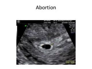
Ultrasonography can help distinguish these two groups if retained products are visualized in the uterine cavity .
Any physical or sonographic evidence of retained products of conception should prompt consideration of suction curettage to complete evacuation of the uterus. • In the absence of detectable retained material, a presumptive diagnosis of endometritis may be made and treated with a trial of broad spectrum antibiotic therapy, with coverage of anaerobes (eg, cefotetan [2 g intravenously] plus doxycycline [100 mg intravenously or orally] every 12 hours). This regimen can be completed as an outpatient oral regimen for a 14-day course.
Generalized abdominal tenderness, • guarding, • tachycardia, • high fever, and prostration • suggest advanced sepsis. • An alternative outpatient regimen is ceftriaxone 250 mg intramuscularly in a single dose plus doxycycline 100 mg orally twice a day for 14 days with or without metronidazole 500 mg orally twice a day for 14 days. • >>> These patients require aggressive therapy with broad spectrum intravenousantibiotics, uterine re-evacuation, assessment for uterine perforation, and monitoring and support in an intensive care unit.
Ongoing pregnancy — Direct or indirect injury to the developing embryo could occur. increased risk of Moebiussequence with autism in children exposed to misoprostol in the first trimester Moebius sequence is a clinical condition characterized by ophthalmic-facial palsy and muscle or bone malformations in the limbs. It represents a cascade of events resulting from embryo trauma from varied etiologies (eg, genetic factors, environmental injuries, prolonged membrane ruptures and chorion villus sampling). is more likely to be a complication of early rather than late abortion. Return of the serum hCG concentration to undetectable following pregnancy termination varies widely from 7 to 60 days The hCG concentration peaks at 8 to 11 weeks at approximately 90,000 mIU. The decline in serum hCG is rapid for the first several days (half-life 9 to 31 hours) and then proceeds more slowly (half-life 55 to 64 hours) An ongoing intrauterine pregnancy may occur after an attempted pregnancy termination if the products of conception are not closely examined by an experienced clinician at the time of the procedure (and by a pathologist) to verify successful completion. Alternatively, ongoing pregnancy may rarely result from a multiple gestation in which only one of the sacs was aborted. reported an ongoing pregnancy rate of 1.3 per 1000 procedures for pregnancies less than 6 weeks of gestation
Future pregnancies risk of bearing low birth weight babies, delivering prematurely, or suffering spontaneous abortions in subsequent pregnancies in women who carried their first pregnancy to term versuswhose first pregnancy ended in induced abortion is controversy in different studies. All of these retrospective studies are flawed because they are subject to recall bias and inadequate adjustment of other risk factors for adverse pregnancy outcome (eg, sexually transmitted disease, smoking).
CONTRACEPTION — Contraception should be discussed prior to and reviewed immediately after the termination. Ovulation can occur two weeks after a first trimester abortion, thus immediate contraception is important (if desired) and can be initiated the day of the procedure. • A routine follow-up visit two to four weeks after the procedure is intended to confirm that the abortion is complete and to diagnose and treat complications. • Pregnancy symptoms generally should have dissipated within one week of the pregnancy termination and normal menses should return within six weeks. • suggest : • ongoing intrauterine or • ectopic pregnancy or • incomplete abortion
Fever, • pelvic pain, • uterine enlargement, • heavy bleeding, • passage of tissue, • and on-going pregnancy symptoms • require further evaluation.
Any sexually active female is at risk for sexually transmitted infection (STI) associated (PID)
Pelvic inflammatory disease (PID) • acuteand subclinicalinfection of the upper genital tract: • uterus, fallopian tubes, and ovaries; • (endometritis, salpingitis, oophoritis, peritonitis, peri-hepatitis, and/or tubo-ovarian abscess)
Clinicaldiagnosis remains the most important practical approach.
Rare case: • PID can occur in the first 12 weeks of gestation of pregnancy before mucus plug formation. instrumentationof the cervix (eg, termination of pregnancy) are at higher risk • Olderwomen less commonly present with PID, but the cause is more likely to be non-STI related. • Salpingitis in premenarchealgirls and adolescents who are not sexually active, is very rare. respiratory and enteric bacteria should be also considered.
Acute symptomatic PID • Acute onset of • lower abdominal or pelvic pain, • pelvic organ tenderness, • and evidence of inflammation of the genital tract. • The findings can be subtle and nonspecific.
Lower abdominal pain is the (cardinalpresentingsymptom) • usuallybilateralpain and rarely of more than twoweeks' duration • May be quite subtle. • May worsens during coitus or with jarringmovement • May start during or shortly after menses(particularly suggestive ) • May with Abnormal uterine bleeding(post-coital bleeding, inter-menstrual bleeding, menorrhagia) • May with urinary frequency and abnormal vaginal discharge.
physical examination, • abdominaltendernessin the lower quadrants, which may or may not be symmetrical. • Acute cervical motion, uterine, and adnexal tenderness on bimanual pelvic (characteristic of acute PID) • Purulent endocervicaldischarge and/or vaginal discharge is also common.
Rebound tenderness, • fever, • decreased bowel sounds Showed more severe PID. • significant lateralization of adnexal tenderness is uncommon in PID.
Laboratory — • are nonspecific. • peripheral blood leukocytosis In more severe disease • elevated erythrocyte sedimentation rate (ESR) and C-reactive protein (CRP) have poor sensitivity and specificity.
PID should be suspected inThe index of suspicion for PID should be high any young or sexually active female presents with lower abdominal pain and pelvic discomfort.
An indolent presentation of PID with low-gradefever, weight loss, and abdominal pain has been reported with actinomycosis(IUD) and tuberculosis.
clinical diagnosis of PID can be made : • on the basis of history and physicalexamfindingsalone. • laboratory testing** is also done at the initial evaluation of all patients with suspected PID, • empiric treatment should not be delayed
The following tests should be performed for all women suspected of having PID: • Pregnancy test • Microscopyof vaginal discharge • Nucleic acid amplification tests (NAATs) for C.trachomatis and Neisseriagonorrhoeae ,Mycoplasmagenitalium • HIVscreening • Serologic testing for syphilis • Urinalysis is also checked in women with urinary symptoms. • Hepatitis B virus testing ( depending on the patient's riskhistory )
A complete blood count, erythrocyte sedimentation rate, and C-reactive protein are often obtained in who have more severe clinical presentations, including fever.
Ultrasoundis generally the preferred imaging modality if an abscess or adnexal pathology is suspected clinically • for women who are acutely ill (eg, with fever, peritonitis, or a pelvic mass), • whose symptoms are atypical (eg, with an abnormal site or duration of symptoms) or • do not improve significantly within 72 hours after starting empiric antibiotic therapy, or who have persisting pain after completing therapy.
Absenceof radiographic findings consistent with PID does not rule out the possibility of PID and should not be a reason to forgo or delay therapy for presumptive PID.
sonographic findings • operator-dependent, and • often minimal changes seen in women with uncomplicated PID. • Thickened, fluid-filled fallopian tubes, the cogwheel sign (on a cross-section of the tube) may be present. • fluid or gas within the endometrial canal, heterogeneous thickening, or indistinctness of the endometrial stripe, Among women who have endometritis, • a complex thick-walled, multilocular cystic collection in the adnexa, typically with internal echoes or multiple fluid levels, Whena tubo-ovarian abscess is present,
Laparoscopy • can be a useful part of the diagnostic workup for PID when imaging studies have not been definitively informative • In a patient who has failed outpatient treatment for PID, • In a patient wo not clearly improving or worsening after approximately 72 hours of inpatient treatment for PID, • some surgeons may proceed directly to laparoscopy in an acutely ill patient with a high suspicion of a competing diagnosis that would be diagnosedand intervenedon through laparoscopy (eg, appendicitis).
DIAGNOSIS clinical diagnosis • of PID is made • in sexually active young women, • who present with pelvic or lower abdominal pain • and have evidence of cervical motion, uterine, or adnexal tenderness on exam.
The sensitivity of this clinical diagnosis is only 65 to 90% but because of the potential for serious reproductive sequelae if PID treatment is delayed or not given, this presumptive diagnosis is sufficient to warrant empiric antimicrobial therapy for PID. Even patients with minimal or subtle findings should be treated since the potential consequences of withholding therapy are great!
additional findings used tosupportthe clinical diagnosis of PID • Oraltemperature >101°F (>38.3°C) • ● Abnormal cervical or vaginal mucopurulentdischargeor cervical friability • ● Presence of (WBCs) on saline microscopy of vaginal secretions (eg, >15 to 20 WBCs per high power field or more WBCs than epithelial cells) • The CDC also lists an elevated C-reactive protein (CRP) or erythrocyte sedimentation rate (ESR) • ● Documentation of cervical infection with N. gonorrhoeae or C. trachomatis
Certain findings can suggest against PID, such as : • the combination of normal cervical discharge and absence of WBCs on microscopy of vaginal secretions. • prominent gastrointestinal and urinary symptoms can suggest other etiologies of pelvic pain.
certain findings can help to confirm the diagnosis of PID, although their absence does not rule out the possibility of PID: • Pelvic imaging (thickened, fluid-filled tubes/oviducts with or without free pelvic fluid, or tuboovariancomplex) • Doppler studies (tubal hyperemia) • Laparoscopic abnormalities(tubal erythema, edema, and adhesions; purulent exudate or cul-de-sac fluid; and abnormal fimbriae.) • Histologic evidence of endometritis in a biopsy.
Standards for the diagnosis of subclinical PID remain to be established.