Download
1 / 31
370 likes | 793 Views
Dr. Altdorfer: The tongue – Anatomy, histology, innervation. Sulcus terminalis. Foramen cecum. Root. Follicular part. Dorsum linguae. Papillar part. Apex linguae. Intrinsic muscles Sup. longitudinal Inf. longitudinal vertical transversal. SUBLINGUAL REGION. frenulum linguae

E N D
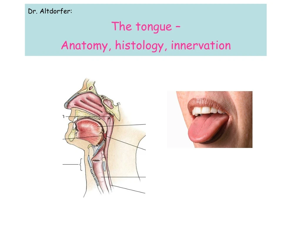
Dr. Altdorfer: The tongue – Anatomy, histology, innervation
Sulcus terminalis Foramen cecum Root Follicular part Dorsum linguae Papillar part Apex linguae
Intrinsic muscles • Sup. longitudinal • Inf. longitudinal • vertical • transversal
SUBLINGUAL REGION • frenulumlinguae • deeplingualvein • sublingual fold • sublingualcaruncula (papilla)
MEDIAL + LATERAL LINGUAL GROOVES Hyoglossus m. Genioglossus Mylohyoid m. Lingual a. Submandibular gland Hypoglossal n.
CT 1. M. mentalis 2. M. depressor angulis oris 3. Mandibula 4. M. mylohyoideus 5. V. sublingualis, Sulcus lateralis linguae 6. M. genioglossus 7. M. hyoglossus 8. Gl. submandibularis 9. V. facialis 10. A. carotis ext. 11. Oropharynx 12. A. facialis 13. V. facialis communis 14. Platysma 15. V. jugularis int. 16. M. stylohyoideus 17. A. carotis int. 18. Mm. longi capitis et colli 19. M. sternocleidomast. 20. V. jugularis ext. 21. M. levator scapulae 22. A. cervic. prof. 23. M. semispin. cerv. 24. M. semispin. capit. 25. M. splenius capitis 26. M. trapezius
Csillag A, Anatomy of the Living Human, Könemann, Cologne, 1999 CT 19. A. vertebralis 20. Spatium retropharyngeum 21. Mm. longi capitis et colli 22. M. scalenus post. 23. M. levator scapulae 24. M. splenius capitis 25. M. trapezius 26. Mm. longissimi et semispin. capit. 27. M. semispinalis cervicis 28. Lig. nuchae 1. Mandibula 2. M. mylohyoideus 3. M. hyoglossus 4. M. genioglossus 5. A. lingualis 6. Gl. submandibularis 7. Platysma 8. V. facialis 9. A. carotis ext. 10. Oropharynx 11. Os hyoideum 12. Cart. thyroidea, cornu sup. 13. A. carotis int. 14. V. jugularis int. 15. M. sternocleidomastoideus 16. V. jugularis ext. 17. M. scalenus ant. 18. M. scalenus medius
Blood supply Lingual a. (ECA) • dorsal lingual a. • sublingual a. • deep lingual a. • Tonsillary branches! Veins • dorsal lingual v. • sublingual v. • deep lingual v. facial vein, retromandibular vein int. jugular vein
Lymphatics: - APEX SUBMENTAL lymph nodes • DORSUM LINGUE SUBMANDIBULAR lymph nodes • ROOT of TONGUE DEEP CERVICAL lymph nodes
Innervation Motor XII. Taste VII. & IX. GSS V., IX.
Papillae filiform fungiform foliate vallate
Circumvallate papilla Tastebud
Glands of the tongue Nuhn-Blandin von Ebner Weber mucous serous mixed
Literature: • Anatomy, Histology atlases • Dr. Gallatz’s lecture images