Download
1 / 38
390 likes | 930 Views
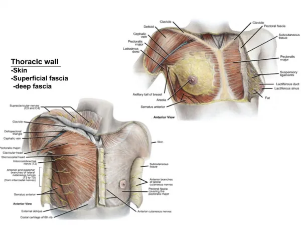
FASCIA, MUSCLES, TENDONS. Skeletal Muscle Structure. Origin: Proximal attachment Insertion: Distal attachment Tendons: Peritendineum Aponeurosis . Skeletal Muscle Hierarchy. Myofilament: Thick filaments Myosin Thin filaments Actin Myofibril:

E N D
Skeletal Muscle Structure • Origin: Proximal attachment • Insertion: Distal attachment • Tendons: Peritendineum • Aponeurosis
Skeletal Muscle Hierarchy • Myofilament: Thick filaments Myosin Thin filaments Actin • Myofibril: Bundle of myofilaments Segmentally arranged into sarcomeres
Skeletal Muscle Hierarchy • Myofiber Made up of many myofibrils Multinucleated in skeletal muscles = muscle cell • Fascicle Bundle of myofibers • Muscle Composed of several to several hundred fascicles
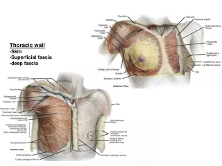
Connective Tissues • Endomysium: Surrounds a myofiber • Perimysium: Surrounds a fascicle
Connective Tissues • Epimysium: Covers entire muscle Blends in with deep fascia • Connective tissue supports provide physical support and a pathway for nerves and vessels.
Myofilaments • Actin myofilaments (F-actin): Polymers of G-actin Tropomyosin Troponin • Myosin filaments: ATPase
Sarcomeres • Z-line (Z-actin): Composed of Z-actin Marks ends of Z-actin • I bands: Part of a sarcomere composed entirely of actin
Sarcomeres • A band: Part of a sarcomere composed of both actin and myosin • H band: Part of a sarcomere composed entirely of myosin
Sliding Filament Theory • During a contraction: I band and H band shorten A band remains the same length • Sliding occurs when ATPase heads of myosin attach to actin via troponin and swivel.
Myofiber • Sarcoplasmic reticulum: Equivalent to endoplasmic reticulum of most cells. • T-tubules: Tubular extensions of the muscle fiber membrane that extend down into the cytoplasm (saracoplasm). Conduct action potential from cell membrane surface to interior.
Myofiber • Cisternae: Saccular extensions of the sarcoplasmic reticulum that release calcium ions in response to action potential. Calcium ions trigger sliding of myosin and actin filaments, resulting in a contraction.
Myofiber Type • The myofiber type (red or white) depends on innervation. • All myofibers in a motor unit are of the same type.
Dark (red) Fibers • Fatigue resistant • Contract slowly (slow twitch) • Rely on oxidative phosphorylation • Have a large number of mitochondria • Have a high concentration of myoglobin • Have a low concentration of ATPase
Light (white) Fibers • Fatigue easily • Contract rapidly (fast twitch) • Rely on glycolysis • Have a small number of mitochondria • Have a low concentration of myoglobin • Have a high concentration of ATPase
Neuromuscular Junctions • Components: Presynaptic membrane: Terminal end of motor neuron. Synaptic cleft Postsynaptic membrane: Sarcolemma (cell membrane of myofiber)
Motor Unit • Consists of a motor neuron and all the myofibers it innervates Units for fine control have fewer fibers Units for gross control have many fibers
Muscle Classification • Fiber arrangement • Shape • Origin and insertion • Function
Fiber Arrangement • Straight: Example: rectus abdominis • Fusiform: Example: biceps brachii • Unipennate: Example: palmar interosseous muscles • Bipennate: Example: dorsal interosseous muscles • Multipennate: Example: deltoid muscle
Muscle Shape • Deltoid • Trapezius
Muscle Origin/Insertion • Coracobrachialis • Sternocleidomastoid
Muscle Function • Pronater teres • Extensor digitorum
Contraction • Definition: A contraction is a muscle’s response to a stimulus. • Types of contraction: Isotonic (the length of the muscle changes) Concentric (length decreases) Eccentric (length increases) Isometric (the length of the muscle stays the same)
Types of Action • Agonist (prime mover): A muscle that primarily carries out the desired action. • Antagonist: A muscle that opposes the agonist. • Synergist: A muscle that eliminates the unwanted action of the agonist.
Types of Action • Fixator: A muscle that stabilizes the origin of another muscle. • Note: a single muscle can be all the above at one time or another.
Insufficiency • Refers to the inability of a multijoint muscle to maximally contract simultaneously over all joints crossed. Active: Refers to the agonist Passive: Refers to the antagonist
Smooth Muscle • Synonyms: Visceral Involuntary • Found in: Walls of visceral tubes (intestines, etc.) Associated with hair follicles Around glandular structures In walls of blood vessels
Smooth Muscle Characteristics • Bundles of sheets of individual cells. • Not striated (smooth). • Cells are primarily elongated and tapered. • Mononucleated.
Smooth Muscle Characteristics • Nuclei are centrally located in each cell. • Does not conduct action potential. • Cells connected by gap junctions. • Not under voluntary control.
Cardiac Muscle Tissue • Found only in walls of heart. • Characteristics: Striated (sarcomeres) Mononucleated cells: May branch Intercalated discs: Sites of transfer of stimulus between adjacent cells.