Download
1 / 30
300 likes | 463 Views
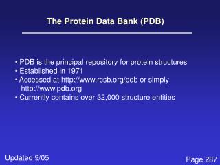
Validation and Deposition at the RCSB Protein Data Bank. Or, how to make your life (and mine) easier. Kyle Burkhardt, Lead Data Annotator The RCSB PDB at Rutgers University. www.pdb.org deposit@rcsb.rutgers.edu. 5 Easy Steps for Fast, Accurate, and Complete Data Deposition.

E N D
Validation and Deposition at the RCSB Protein Data Bank Or, how to make your life (and mine) easier Kyle Burkhardt, Lead Data Annotator The RCSB PDB at Rutgers University www.pdb.org deposit@rcsb.rutgers.edu
5 Easy Steps for Fast, Accurate, and Complete Data Deposition • Use pdb_extract • Validate your entry • Verify sequence • Use Ligand Depot • Deposit with ADIT This is an iterative process
1.Use • RCSB pdb_extract • Extracts data from crystal structure determination programs • Fills in many fields automatically • Allows input of additional information • Generates a complete data file ready for deposition
Availability • CCP4 package (CCP4i interface) • Script and command line • Desktop (script and command line) • deposit.pdb.org/mmcif/PDB_EXTRACT • Web-based • pdb-extract.rutgers.edu • Tutorial • pdb-extract.rutgers.edu/tutorial.html
2.Validate Your Entry • RCSB PDB Validation Server • Reads mmCIF file from pdb_extract • Reads PDB or mmCIF files from refinement programs • Reads structure factor file in mmCIF format • Steps in Validation 1. Precheck coordinate and experimental data files 2. Produce validation report
Validation Reports Contain: • Close contacts • Bond and angle deviations • Chirality errors • Sequence/coordinate (mis)alignment • Missing and extra atoms or residues • Distant waters • NUCheck1, PROCHECK2, and SFCHECK3 1. Feng Z, Westbrook J, Berman HM.(1998) NUCheck: Rutgers University, New Brunswick, NJ. Report No.: NDB-407. 2. Laskowski, R.A., McArthur, M.W., Moss, D.S., et al. (1993) PROCHECK: a program to check the stereochemical quality of protein structures. J. Appl. Cryst. 26:283-291. 3. Vaguine A.A., Richelle J., Wodak S.J. (1999) SFCHECK: a unified set of procedures for evaluating the quality of macromolecular structure-factor data and their agreement with the atomic model. Acta Crystallogr. D55:191-205.
Jul. 10, 15:47:11 2004 Thank you for using RCSB. The following geometrical and stereochemical features have been calculated for your structure: CLOSE CONTACTS -------------- ==> Close contacts in same asymmetric unit. Distances smaller than 2.2 Angstroms are considered as close contacts. Chain Atom Res Seq Chain Atom Res Seq Symm_Code Distance ------------------------------------------------------------------------- L O PRO 56 - O HOH 235 ( 1, 5, 5, 5) Dist = 1.52 M NE ARG 244 - O HOH 417 ( 1, 5, 5, 5) Dist = 1.67 L O LYS 48 - L OE2 GLU 57 ( 1, 5, 5, 5) Dist = 1.67 L C PRO 56 - O HOH 235 ( 1, 5, 5, 5) Dist = 1.67 E CB LYS 54 - E O LEU 66 ( 1, 5, 5, 5) Dist = 1.69 M O TRP 242 - O HOH 347 ( 1, 5, 5, 5) Dist = 1.71 L OG SER 99 - O HOH 464 ( 1, 5, 5, 5) Dist = 1.73 ==> Close contacts based on crystal symmetry. Distances smaller than 2.2 Angstroms are considered as close contacts. Chain Atom Res Seq Chain Atom Res Seq Symm_Code Distance ------------------------------------------------------------------------- E OG1 THR 198 - H NE ARG 108 ( 2, 6, 4, 5) Dist = 1.92 E O THR 198 - H NH1 ARG 169 ( 2, 6, 4, 5) Dist = 2.03 H NE2 GLN 226 - I NZ LYS 75 ( 2, 5, 4, 6) Dist = 2.15
Validation Availability • Desktop • deposit.pdb.org/mmcif/VAL • Web-based • deposit.pdb.org/validate • pdb_extract • Command line option • ADIT • Desktop and Web • Tutorial • deposit.pdb.org/validate/docs/tutorial.html
3.Verify Sequence • Input the complete deposition sequence (e.g. BLAST www.ncbi.nih.gov/BLAST1) • Include • residues missing due to lack of electron density • cloning artifacts and HIS tags that were not cleaved • mutations or substitutions • Output compares the deposition sequence to sequence database references. • Check sequence database correspondence 1.Altschul, S.F., Gish, W., Miller, W., Myers, E.W. & Lipman, D.J. (1990) "Basic local alignment search tool." J. Mol. Biol. 215:403-410
Sequence Discrepancies • Alanine or glycine mismatches • GLU/GLN or ASP/ASN mismatches • Intended mutation • Deletion or insertion • Unobserved gap • Real or unexpected difference • “We’re right and they’re wrong”
Sample BLAST output >gi|126605|sp|P00720|LYCV_BPT4 Lysozyme (Lysis protein) (Muramidase) (Endolysin) Length = 164 Score = 189 bits (440), Expect = 2e-48 Identities = 65/80 (81%), Positives = 67/80 (83%), Gaps = 6/80 (7%) Query: 1 MNIFEMLRIDQGLAAAAAANTEGYYTIGIGHLLT------AAKSELDKAIGRNTNGVITK 54 MNIFEMLRID+GL +TEGYYTIGIGHLLT AAKSELDKAIGRN NGVITK Sbjct: 1 MNIFEMLRIDEGLRLKIYKDTEGYYTIGIGHLLTKSPSLNAAKSELDKAIGRNCNGVITK 60
4.Use • RCSB PDB Ligand Depot • Use to find code for existing ligands • Searching by many attributes • New ligands • E-mail chemical diagram (with bond order), IUPAC name, synonyms, and formula to deposit@rcsb.rutgers.edu • Choose your three letter code for new ligands • Access • ligand-depot.rutgers.edu
3-letter code name, formula synonyms diagram PDB entries listed by resolution coordinate download
5.Deposit with • Web-based ADIT (deposit.pdb.org/adit/) • Load file (coordinates and sfs) • Input missing information • Deposit • Desktop ADIT (deposit.pdb.org/mmcif/ADIT) • Load file (coordinates and sfs), add missing information, validate and save • Deposit • Load in Web-based ADIT and deposit Tutorial deposit.pdb.org/adit/docs/tutorial.html
Important Points to Consider • Title • Sequence (including mutations) • Protein name • Biological unit • Ligands • Visual inspection of the entry • Unusual situations Don’t be shy. Talk is good. Tell us the whole story right away.
I just deposited. Now what happens? • What is annotation? • “A note added by way of comment or explanation” • When do we annotate? • All the time! You never stop depositing • Where do we annotate? • RCSB-PDB @ Rutgers and Prague (don’t ask) • Who else annotates? MSD/EBI, PDBj • Why do we annotate? • Annotators are here to help you represent your data in the best possible way • How do we annotate? • We use the same tools we want you to use
What Do Annotators Do? • Annotators check everything • Check entry for self-consistency • Check title • Check citation references with PubMed (http://pubmed.gov/) • Correct format errors in data and coordinates • Check sequence • Add sequence database reference • Add protein name and synonyms • Check source • Check ligand nomenclature • Add biological unit information • Visually check entry • Generate validation reports
After Initial Annotation • Correspond with you • Update entries • Corrections, new coordinate sets • Release entries • How long does the entire process take? • Annotation is like a box of chocolates…
Release Information • Release options • Pre-release of sequence • Coordinate release • Release immediately • Hold until publication (HPUB) • Hold until a particular date • HPUB and HOLD limit • not more than 1 year after deposition • It’s ok to release a structure without a citation
How Do We Find Citations? • Some journals • PDB users • Weekly PubMed searches • You tell us (please tell us!)
How to make my life more difficult • Deposit before you finish your refinement • Don’t use any software tools • Don’t follow deposition procedures • Deposit the day you leave your company or postdoc position • Deposit the day the journal needs the PDB ID • Don’t tell us the whole story until later • Provide only one contact author • Don’t tell us when the structure has been published • Don’t acknowledge our e-mail It makes your life more difficult too…
Why Should You Do What I Say? • Create a more complete deposition with less manual input • Minimize mistakes • Check (and recheck) • Give yourself time to deposit • Save time (for you and us) • Help us help you!
Please Visit the RCSB PDB Booth #325 in “Data Alley” • Demonstrations • pdb_extract • validation • ADIT • reengineered PDB site demos during coffee breaks • Questions answered • Tattoos, posters and literature • Poster session Tuesday night #P142 You can always write to us at deposit@rcsb.rutgers.edu All information is available from deposit.pdb.org
Our friendly annotation staff thanks you! Top row: Suzanne Richman, Shri Jain, Jasmin Yang, Bohdan Schneider, Kyle Burkhardt, Shuchismita Dutta, Zukang Feng. Bottom row: Lew-Christiane Fernandez, Irina Persikova, Alice Xenachis. Other RCSB-Rutgers members: Helen M. Berman, Li Chen, Judith L. Flippen-Anderson, Vladimir Guranovic, Susan van Arnum, John Westbrook, Huanwang Yang, Christine Zardecki
Acknowledgements • The Protein Data Bank (PDB) is operated by • Rutgers, The State University of New Jersey • San Diego Supercomputer Center at the University of California, San Diego • Center for Advanced Research in Biotechnology/UMBI/NIST • The RCSB PDB is supported by funds from • National Science Foundation (NSF) • National Institute of General Medical Sciences (NIGMS) • Office of Science, Department of Energy (DOE) • National Library of Medicine (NLM) • National Cancer Institute (NCI) • National Center for Research Resources (NCRR) • National Institute of Biomedical Imaging and Bioengineering (NIBIB) • National Institute of Neurological Disorders and Stroke (NINDS) • The worldwide PDB (wwPDB) is a collaboration between • RCSB • MSD/EBI • PDBj
RCSB-PDB Data Deposition Services • pdb_extract • Web- http://pdb-extract.rutgers.edu/ • Standalone - http://deposit.pdb.org/mmcif/PDB_EXTRACT/index.html • Validation Server • Web - http://deposit.pdb.org/validate/ • Standalone - http://deposit.pdb.org/mmcif/VAL/index.html • ADIT • Web – http://deposit.pdb.org/adit/ • Standalone - http://deposit.pdb.org/mmcif/ADIT/index.html • Ligand Depot - http://ligand-depot.rutgers.edu/ • Overview and tutorials for all RCSB-PDB data deposition services – http://deposit.pdb.org