Download
1 / 68
930 likes | 1.69k Views
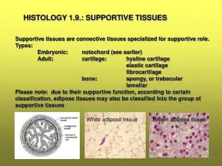
Histology (Tissues). Anatomy & Physiology I Chapter 4. Histology. Histology - study of tissues Tissue - a collection of similar cells that group together to perform a specialized function. Four main groups of tissues Epithelial Connective Muscle Nervous tissue.

E N D
Histology (Tissues) Anatomy & Physiology I Chapter 4
Histology • Histology - study of tissues • Tissue - a collection of similar cells that group together to perform a specialized function. • Four main groups of tissues • Epithelial • Connective • Muscle • Nervous tissue Tissue Classification
Tissue Classification Nervous tissue:Internal communication • Brain, spinal cord, and nerves Muscle tissue:Contracts to cause movement • Muscles attached to bones (skeletal) • Muscles of heart (cardiac) • Muscles of walls of hollow organs (smooth) Epithelial tissue:Forms boundaries between different environments, protects, secretes, absorbs, filters • Skin surface (epidermis) • Lining of GI tract organs and other hollow organs Connective tissue:Supports, protects, binds other tissues together • Bones • Tendons • Fat and other soft padding tissue
Epithelial Tissue • Forms a protective covering for the body • Is the main tissue of outer layer of skin • Forms membranes, ducts, and the lining of body cavities and hollow organs • Layers of closely adhering cells • Flat sheet with upper surface exposed to the environment or an internal body cavity • No blood vessels - underlying connective tissue supplies oxygen • Rests on basement membrane - anchors epithelium to connective tissue
Structure of Epithelial Tissue Classification by shape • Squamous – flat cells • Cuboidal – cube shaped cells • Columnar – tall, narrow cells Classification by arrangement • Simple – one layer of cells; all cell in contact with basement membrane • Stratified – two or more layers of cells • Pseudostratified – one layer of cells but appears to be more
Simple vs. Stratified Epithelia • Simple epithelium • allows diffusion and secretion of materials • Stratified epithelium • protection in areas subject to wear and tear • Classes of • epithelium Pseudostratified columnar Simple Stratified • Cell shapes Squamous Cuboidal Columnar
Epithelial Tissue (Epithelium) Two main types (by location): • Covering and lining epithelia • On external and internal surfaces • Glandular epithelia • Secretory tissue in glands
Overview of Epithelial Tissues • For each of the following types of epithelia, note: • Description • Function • Location
Simple Squamous Epithelium Description: Single layer of flattened cells with disc-shaped central nuclei and sparse cytoplasm; the simplest of the epithelia. Air sacs of lung tissue Function: Allows passage of materials by diffusion and filtration in sites where protection is not important; secretes lubricating substances in serosae. Nuclei of squamous epithelial cells Location: Kidney glomeruli; air sacs of lungs; lining of heart, blood vessels, and lymphatic vessels; lining of ventral body cavity (serosae). Photomicrograph: Simple squamous epithelium forming part of the alveolar (air sac) walls (125x).
Epithelia: Simple Squamous • Two other locations • Endothelium • The lining of lymphatic vessels, blood vessels, and heart • Mesothelium • The epithelium of serous membranes in the ventral body cavity
Simple Cuboidal Epithelium Description: Single layer of cubelike cells with large, spherical central nuclei. Simple cuboidal epithelial cells Function: Secretion and absorption. Basement membrane Location: Kidney tubules; ducts and secretory portions of small glands; ovary surface. Connective tissue Photomicrograph: Simple cuboidal epithelium in kidney tubules (430x).
Simple Columnar Epithelium Description: Single layer of tall cells with round to oval nuclei; some cells bear cilia; layer may contain mucus- secreting unicellular glands (goblet cells). Simple columnar epithelial cell Function: Absorption; secretion of mucus, enzymes, and other substances; ciliated type propels mucus (or reproductive cells) by ciliary action. Location: Nonciliated type lines most of the digestive tract (stomach to anal canal), gallbladder, and excretory ducts of some glands; ciliated variety lines small bronchi, uterine tubes, and some regions of the uterus. Basement membrane Photomicrograph: Simple columnar epithelium of the stomach mucosa (860X).
Pseudostratified Columnar Epithelium Description: Single layer of cells of differing heights, some not reaching the free surface; nuclei seen at different levels; may contain mucus- secreting cells and bear cilia. Cilia Mucus of mucous cell Pseudo- stratified epithelial layer Function: Secretion, particularly of mucus; propulsion of mucus by ciliary action. Location: Nonciliated type in male’s sperm-carrying ducts and ducts of large glands; ciliated variety lines the trachea, most of the upper respiratory tract. Basement membrane Photomicrograph: Pseudostratified ciliated columnar epithelium lining the human trachea (570x). Trachea
Stratified Squamous Epithelium Description: Thick membrane composed of several cell layers; basal cells are cuboidal or columnar and metabolically active; surface cells are flattened (squamous); in the keratinized type, the surface cells are full of keratin and dead; basal cells are active in mitosis and produce the cells of the more superficial layers. Stratified squamous epithelium Function: Protects underlying tissues in areas subjected to abrasion. Nuclei Location: Nonkeratinized type forms the moist linings of the esophagus, mouth, and vagina; keratinized variety forms the epidermis of the skin, a dry membrane. Basement membrane Connective tissue Photomicrograph: Stratified squamous epithelium lining the esophagus (285x).
Stratified Epithelia • Stratified epithelium is found in tissues subject to wear and tear; provides protection to underlying tissues; retards water loss through skin; resists penetration by pathogenic organisms • range from 2 to 20 or more layers of cells • some cells resting directly on others; only the deepest layer attaches to the basement membrane • three stratified epithelia are named for the shapes of their surface cells • most widespread epithelium in the body • deepest layers undergo continuous mitosis • their daughter cells push toward the surface and become flatter as they migrate farther upward • finally die and flake off – exfoliation or desquamation
Epithelia: Stratified Cuboidal • Quite rare in body • Found in some sweat and mammary glands • Typically two cell layers thick
Epithelia: Stratified Columnar • Limited distribution in body • Small amounts in pharynx, male urethra, and lining some glandular ducts • Also occurs at transition areas between two other types of epithelia
Transitional epithelium Description: Resembles both stratified squamous and stratified cuboidal; basal cells cuboidal or columnar; surface cells dome shaped or squamouslike, depending on degree of organ stretch. Transitional epithelium Function: Stretches readily and permits distension of urinary organ by contained urine. Location: Lines the ureters, urinary bladder, and part of the urethra. Basement membrane Connective tissue Photomicrograph: Transitional epithelium lining the urinary bladder, relaxed state (360X); note the bulbous, or rounded, appearance of the cells at the surface; these cells flatten and become elongated when the bladder is filled with urine.
Glandular Epithelia • A gland is an organ specialized to produce a substance for use elsewhere in the body or releases them for elimination from the body • Classified by: • Site of product release—endocrine or exocrine • Relative number of cells forming the gland—unicellular (e.g., goblet cells) or multicellular
Endocrine and Exocrine Glands • Glands are composed of epithelial tissue in a connective tissue framework and capsule • exocrine glands - maintain their contact with the body surface by way of a duct (epithelial tube that conveys secretion to surface) • sweat, mammary and tear glands • endocrine glands - lose their contact with the surface and have no ducts • hormones – secretion of endocrine glands • secrete (hormones) directly into blood • thyroid, adrenal and pituitary glands • some organs have both endocrine and exocrine function (liver, gonads, pancreas)
Exocrine Glands • More numerous than endocrine glands • Secrete products into ducts • Secretions released onto body surfaces (skin) or into body cavities • Examples: • mucous glands • sweat glands • Oil glands • salivary glands
Exocrine Glands • Single cell • Goblet cell (secretes mucus) • The only important unicellular gland is the goblet cell • Multiple cells • Simple Tubular (found in intestine) • Branched tubular (found in stomach) • Coiled tubular (sweat glands) • Saclike or alveolar (sebaceous glands) • Compound (salivary glands)
Microvilli Secretory vesicles containing mucin Rough ER Golgi apparatus Nucleus
Multicellular Exocrine Glands • Multicellular exocrine glands are composed of a duct and a secretory unit • Classified according to: • Duct type (simple or compound) • Structure of their secretory units (tubular, alveolar, or tubuloalveolar)
Types of Exocrine Glands Simple coiled tubular Compound areolar Compound tubuloareolar • simple - unbranched duct • compound - branched duct • shape of gland • tubular – duct and secretory portion have uniform diameter • alveolar - secretory cells form dilated sac (alveolar or sacklike ) • tubuloalveolar - both tubular and alveolar portions Example: Sweat gland Example: Pancreas Key Example: Mammary gland Duct Secretory unit
Multicellular Exocrine Glands Simple duct structure (duct does not branch) Compound duct structure (duct branches) Tubular secretory structure Simple tubular Simple branched tubular Compound tubular Example Intestinal glands Example Stomach (gastric) glands Example Duodenal glands of small intestine Alveolar (saclike) secretory structure Simple alveolar Simple branched alveolar Compound alveolar Compound tubuloalveolar Example No important example in humans Example Sebaceous (oil) glands Example Mammary glands Example Salivary glands Surface epithelium Duct Secretory epithelium
Methods of Secretion Merocrine Gland • merocrine glands (eccrine glands) – have vesicles that release their secretion by exocytosis • tear glands, pancreas, gastric glands, and others • apocrine glands – primarily merocrine mode of secretion • axillary sweat glands, mammary glands Exocytosis Nucleus Secretory vesicle Merocrine gland
Methods of Secretion Holocrine Gland • holocrine glands – cells accumulate a product and then the entire cell disintegrates • secretion a mixture of cell fragments and synthesized substance • oil glands of scalp, glands of eyelids (b) Holocrine gland
Connective Tissue Characteristics • cells usually occupy less space than the extracellular material • most cells are not in direct contact with each other • separated by extracellular material • highly vascular – richly supplied with blood vessels (except cartilage which is avascular) • most abundant, widely distributed, and histologically variable of the primary tissues
Functions of Connective Tissue • binding of organs – tendons and ligaments • support – bones and cartilage • protection – bones and blood • movement – bones • storage – fat, calcium, phosphorus • transport - blood
Structural Elements of Connective Tissue • Ground substance • Medium through which solutes diffuse between blood capillaries and cells • Components: • Interstitial fluid • Adhesive glycoproteins (“glue”) • Proteoglycans
Connective Tissue Fibers • Collagen (white fibers) • Strongest and most abundant type • tough, flexible, and resist stretching • tendons, ligaments, and deep layer of the skin are mostly collagen • Elastic (yellow fibers) • Thinner than collagen fibers • Allows stretch and recoil • Reticular • thin collagen fibers coated with glycoprotein • form framework of such organs as spleen and lymph nodes
Structural Elements of Connective Tissue • Cells • Immature, secretory cells = “blasts” • Mature cells = “cytes” • Fibroblasts – produce fibers and ground substances • Chondroblasts and chondrocytes in cartilage • Osteoblasts and osteocytes in bone • Hematopoietic stem cells in bone marrow • Adipocytes, leukocytes, mast cells, and macrophages
Cell types Extracellular matrix Ground substance Fibers • Collagen fiber • Elastic fiber • Reticular fiber Macrophage Fibroblast Lymphocyte Fat cell Capillary Mast cell Neutrophil
Connective Tissue Categories Categorized by physical properties • Circulating connective tissue • Blood • Generalized (fibrous) connective tissue • Loose: Areolar, Adipose and Reticular • Dense: Regular, Irregular and Elastic • Structural connective tissue • Cartilage: Hyaline, Elastic, Fibrocartilage • Osseous (Bone) Tissue
Blood Description: Red and white blood cells in a fluid matrix (plasma). Plasma Function: Transport of respiratory gases, nutrients, wastes, and other substances. Neutrophil Location: Contained within blood vessels. Red blood cells Lymphocyte Photomicrograph: Smear of human blood (1860x); two white blood cells (neutrophil in upper left and lymphocyte in lower right) are seen surrounded by red blood cells.
Types of Generalized (Fibrous) Connective Tissue • loose connective tissue • much gel-like ground substance between cells • dense connective tissue • fibers fill spaces between cells Tendons
Areolar (loose connective tissue) Description:Gel-like matrix with all three fiber types; cells: fibroblasts, macrophages, mast cells, and some white blood cells. Elastic fibers Function: Wraps and cushions organs; its macrophages phagocytize bacteria; plays important role in inflammation; holds and conveys tissue fluid. Collagen fibers Location: Widely distributed under epithelia of body, e.g., forms lamina propria of mucous membranes; packages organs; surrounds capillaries. Fibroblast nuclei Epithelium Photomicrograph: Areolar connective tissue, a soft packaging tissue of the body (300x). Lamina propria
Adipose (loose connective tissue) Description: Matrix as in areolar, but very sparse; closely packed adipocytes, or fat cells, have nucleus pushed to the side by large fat droplet. Function: Provides reserve food fuel; insulates against heat loss; supports and protects organs. Nucleus of fat cell Location: Under skin in the hypodermis; around kidneys and eyeballs; within abdomen; in breasts. Vacuole containing fat droplet Adipose tissue Photomicrograph: Adipose tissue from the subcutaneous layer under the skin (350x). Mammary glands
Reticular (loose connective tissue) Description: Network of reticular fibers in a typical loose ground substance; reticular cells lie on the network. Function: Fibers form a soft internal skeleton (stroma) that supports other cell types including white blood cells, mast cells, and macrophages. White blood cell (lymphocyte) Location: Lymphoid organs (lymph nodes, bone marrow, and spleen). Reticular fibers Spleen Photomicrograph: Dark-staining network of reticular connective tissue fibers forming the internal skeleton of the spleen (350x).
Dense Regular Connective tissue Description: Primarily parallel collagen fibers; a few elastic fibers; major cell type is the fibroblast. Collagen fibers Function: Attaches muscles to bones or to muscles; attaches bones to bones; withstands great tensile stress when pulling force is applied in one direction. Location: Tendons, most ligaments, aponeuroses. Nuclei of fibroblasts Shoulder joint Ligament Photomicrograph: Dense regular connective tissue from a tendon (500x). Tendon
Dense Irregular Connective Tissue Description: Primarily irregularly arranged collagen fibers; some elastic fibers; major cell type is the fibroblast. Nuclei of fibroblasts Function: Able to withstand tension exerted in many directions; provides structural strength. Location: Fibrous capsules of organs and of joints; dermis of the skin; submucosa of digestive tract. Collagen fibers Fibrous joint capsule Photomicrograph: Dense irregular connective tissue from the dermis of the skin (400x).
Elastic (dense connective tissue) Description: Dense regular connective tissue containing a high proportion of elastic fibers. Function: Allows recoil of tissue following stretching; maintains pulsatile flow of blood through arteries; aids passive recoil of lungs following inspiration. Elastic fibers Location: Walls of large arteries; within certain ligaments associated with the vertebral column; within the walls of the bronchial tubes. Aorta Photomicrograph: Elastic connective tissue in the wall of the aorta (250x). Heart
Connective Tissue: Cartilage • cartilage lacks blood vessels (avascular) • Cartilage Cells: • Chondroblasts – immature cartilage cells; produce extracellular material (matrix) • Chondrocytes – mature cartilage cells; once trapped in matrix, chondroblasts become chondrocytes • Three types of cartilage: • Hyaline cartilage • Elastic cartilage • Fibrocartilage
Hyaline Cartilage Description: Amorphous but firm matrix; collagen fibers form an imperceptible network; chondroblasts produce the matrix and when mature (chondrocytes) lie in lacunae. Function: Supports and reinforces; has resilient cushioning properties; resists compressive stress. Location: Forms most of the embryonic skeleton; covers the ends of long bones in joint cavities; forms costal cartilages of the ribs; cartilages of the nose, trachea, and larynx. Chondrocyte in lacuna Matrix Costal cartilages Photomicrograph: Hyaline cartilage from the trachea (750x).
Elastic Cartilage Description: Similar to hyaline cartilage, but more elastic fibers in matrix. Function: Maintains the shape of a structure while allowing great flexibility. Chondrocyte in lacuna Location: Supports the external ear (pinna); epiglottis. Matrix Photomicrograph: Elastic cartilage from the human ear pinna; forms the flexible skeleton of the ear (800x).
Fibrocartilage Description: Matrix similar to but less firm than that in hyaline cartilage; thick collagen fibers predominate. Function: Tensile strength with the ability to absorb compressive shock. Location: Intervertebral discs; pubic symphysis; discs of knee joint. Chondrocytes in lacunae Intervertebral discs Collagen fiber Photomicrograph: Fibrocartilage of an intervertebral disc (125x). Special staining produced the blue color seen.
Osseous (Bone) Tissue Description: Hard, calcified matrix containing many collagen fibers; osteocytes lie in lacunae. Very well vascularized. Central canal Function: Bone supports and protects (by enclosing); provides levers for the muscles to act on; stores calcium and other minerals and fat; marrow inside bones is the site for blood cell formation (hematopoiesis). Lacunae Lamella Location: Bones Photomicrograph: Cross-sectional view of bone (125x).
Excitable Tissues: Muscular & Nervous • excitability – ability to respond to stimuli • a characteristic of all living cells but developed to highest degree in nervous and muscular tissues • membrane potential – electrical charge difference (voltage) that occurs across the plasma membranes is the basis for their excitation • respond quickly to outside stimulus by means of changes in membrane potential • nerves – changes result in rapid transmission of signals to other cells • muscles – changes result in contraction, shortening of the cell
Muscular Tissue Functions • muscular tissue is made up of elongated cells that are specialized to contract in response to stimulation • primary job is to exert physical force on other tissues and organs • creates movements involved in body and limb movement, digestion, waste elimination, breathing, speech, and blood circulation • important source of body heat • three types of muscle: skeletal, cardiac, and smooth