Download
1 / 30
300 likes | 389 Views
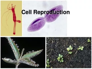
Systems of Cell Reproduction. Four events occur before and during cell division: A signal to reproduce must be received. Replication of DNA and vital cell components must occur. DNA must be distributed to the new cells. The cell membrane or cell wall must separate the two new cells.

E N D
Systems of Cell Reproduction Four events occur before and during cell division: A signal to reproduce must be received. Replication of DNA and vital cell components must occur. DNA must be distributed to the new cells. The cell membrane or cell wall must separate the two new cells.
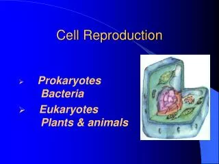
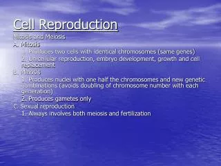
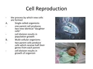
Systems of Cell Reproduction Prokaryotes divide by fission. Most prokaryotes have one circular chromosome. As DNA replicates, each of the two resulting DNA molecules attaches to the plasma membrane. Cytokinesis separates the one cell into two, each with a complete chromosome.
Systems of Cell Reproduction Eukaryotic cells divide by mitosis or meiosis. Eukaryotes usually have many chromosomes. Eukaryotes have a nucleus, which must replicate and, with few exceptions, divide during cell division.
Interphase and the Control of Cell Division The cell cycle has two phases: mitosis and interphase. A typical eukaryotic cell will spend most of its life in interphase, the period between divisions of the cytoplasm. Some cells, such as human nerve and muscle cells, lose the capacity to divide altogether and stay in interphase indefinitely, while other cells divide regularly or occasionally.
Interphase and the Control of Cell Division Interphase consists of three subphases: G1 is Gap 1, the period just after mitosis and before the beginning of DNA synthesis. Next is S phase (synthesis), which is the time when the cell’s DNA is replicated. G2 is the time after S and prior to mitosis.
Interphase and the Control of Cell Division Transitions from G1 to S and G2 to M depend on activation of a protein called cyclin-dependent kinase, or Cdk. A kinase catalyzes phosphorylation of a protein. Cdk is activated by binding to a second type of protein called cyclin. Several different cyclins exist, which, when bound to Cdk, phosphorylate different target proteins.
Figure 9.4 Cyclin-Dependent Kinases and Cyclins Trigger Transisions in the Cell Cycle
Interphase and the Control of Cell Division Cyclin-Cdk complexes act as checkpoints. When functioning properly, they allow or prevent the passage to the next cell cycle stage, depending on the extra- and intracellular conditions. In cancer cells, these cyclin-Cdk controls are often disrupted.
Eukaryotic Chromosomes The basic unit of the eukaryotic chromosome is a gigantic, linear, double-stranded molecule of DNA complexed with many proteins to form a dense material called chromatin. After the DNA of a chromosome replicates during S phase, each chromosome consists of two joined chromatids.
Eukaryotic Chromosomes Interphase chromosomes are wrapped around proteins called histones. These wraps of DNA and histone proteins are called nucleosomes and resemble beads on a string.
Mitosis: Distributing Exact Copies of Genetic Information When the cell enters S phase and DNA is replicated, the centrosome replicates to form two centrosomes. During G2-to-M transition, the two centrosomes separate from each other and move to opposite ends of the nuclear envelope. Centrosomes are regions where microtubules form.
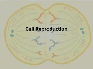
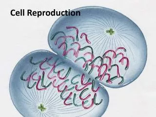
Mitosis: Distributing Exact Copies of Genetic Information Prophase marks the beginning of mitosis. Chromosomes compact and coil, becoming more dense and visible. Spindle forms Kinetochores develop in the region around the centromere.
Mitosis: Distributing Exact Copies of Genetic Information During metaphase, the kinetochores arrive at the equatorial plate. Chromosomes are fully condensed and have distinguishable shapes. Enzymes unravel the interconnected DNA molecules at the centromere, and all the chromatids separate simultaneously.
Mitosis: Distributing Exact Copies of Genetic Information Anaphase begins when the chromatids separate. Move toward poles. Pulled by microtubules which shorten at the poles.
Mitosis: Distributing Exact Copies of Genetic Information Telophase begins when the chromosomes finish moving. Nuclear envelopes and nucleoli coalesce and re-form.
Cytokinesis: The Division of the Cytoplasm Animal cells divide by a furrowing (a “pinching in” or constriction) of the plasma membrane. Microfilaments of actin and the motor protein filament myosin first form a ring beneath the plasma membrane. Actin and myosin contract to produce the constriction.
Cytokinesis: The Division of the Cytoplasm Plants have cell walls and the cytoplasm divides differently. After the spindle breaks down, vesicles from the Golgi apparatus appear in the equatorial region. The vesicles fuse to form a new plasma membrane, and the contents of the vesicles combine to form the cell plate, which is the beginning of the new cell wall. Animation
Meiosis: A Pair of Nuclear Divisions Meiosis consists of two nuclear divisions that reduce the number of chromosomes to the haploid number. The DNA is replicated only once. The functions of meiosis are: To reduce the chromosome number from diploid to haploid. To ensure each gamete gets a complete set of chromosomes. To promote genetic diversity among products.
Meiosis: A Pair of Nuclear Divisions Meiosis I is preceded by an interphase in which DNA is replicated. During prophase I, synapsis occurs: The two homologs are joined together by a complex of proteins. This forms a tetrad, or bivalent, which consists of two homologous chromosomes with two sister chromatids.
Meiosis: A Pair of Nuclear Divisions At a later point, the chromosomes appear to repel each other except at the centromere and at points of attachments, called chiasmata, which appear x-shaped. These chiasmata reflect the exchange of genetic material between homologous chromosomes, a phenomenon called crossing-over. This crossing-over increases genetic variation by reshuffling the genes on the homologs.
Figure 9.16 Crossing Over Forms Genetically Diverse Chromosomes
Meiosis: A Pair of Nuclear Divisions The homologous chromosomes separate in anaphase I. The individual chromosomes are pulled to the poles, with one homolog of a pair going to one pole and the other homolog going to the opposite pole.
Meiosis: A Pair of Nuclear Divisions Meiosis leads to genetic diversity. Synapsis and crossing-over during prophase I mix genetic material of the maternal with that of the paternal homologous chromosomes. Which member of a homologous pair segregates or goes to which daughter cell at anaphase I is a matter of chance. This phenomenon is called independent assortment.
Meiotic Errors Nondisjunction occurs when homologous chromosomes fail to separate during anaphase I, or sister chromatids fail to separate during anaphase II. The result is a condition called aneuploidy. Cells end up with no or extra copies of chromosomes. Failure of chromosome 21 to separate in humans results in trisomy 21—Down syndrome.
Meiotic Errors Polyploids have extra whole sets of chromosomes, and this abnormality in itself does not prevent mitosis. Triploids are 3n; tetraploids are 4n. Although mitosis usually is unimpaired, meiosis is problematic, especially for odd numbers of sets, as in triploidy. Barrier to conception. No fusion of gametes or Zygotes can’t survive.
Cell Death Cells die in one of two ways: necrosis and apoptosis. Necrosis occurs when cells either are damaged by poisons or are starved of essential nutrients. These cells swell and burst.
Cell Death Genetically programmed cell death is called apoptosis: The cell may no longer be needed, e.g., cells of the weblike tissue between the fingers of a developing human fetus. Cells that are old or damaged may need to be replaced. The cell death cycle is controlled by signals. The cell becomes isolated, chops up its own chromatin, and gets ingested by surrounding living cells.