Download
1 / 48
480 likes | 691 Views
Microbial Diseases of the Skin: Staphylococcus and Streptococcus. Skin. Portals of entry: Hair follicles Sweat ducts Sebum ducts Parenteral route. Figure 21.1. For clinical purposes: coagulase-positive v. –negative Correlation between coagulase expression and toxin production

E N D
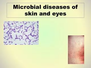
Microbial Diseases of the Skin:Staphylococcus and Streptococcus
Skin Portals of entry: Hair follicles Sweat ducts Sebum ducts Parenteral route Figure 21.1
For clinical purposes: coagulase-positive v. –negative Correlation between coagulase expression and toxin production S. epidermidis Gram-positive cocci, coagulase-negative Up to 90% of normal skin microbiota Only pathogenic when skin barrier is broken Staphylococcus aureus Gram-positive cocci, coagulase-positive Most pathogenic of the staphylococci Various toxins, depending upon infecting strain Staphylococcal Skin Infections
Staphylococcal Skin Infections Common route of entry: skin (hair follicles) Folliculitis: Infection of the hair follicle (often occurs as pimples) Eyelash folliculitis: Sty Folliculitis can progress to an abscess (boil) Abscess: pus surrounded by inflamed tissue Boil can progress to a carbuncle Inflammation of tissue under the skin Always some risk of bacteria entering the bloodstream and producing toxinssepsis
Staphylococcal Skin Infections Sites of infection and diseases caused by Staphylococcus aureus http://textbookofbacteriology.net
Streptococcal Skin Infections Cause wide range of diseases Skin infections are usually localized, but can reach deeper tissue Group A streptococci (GAS): Streptococcus pyogenes Beta-hemolytic streptococci One of the most common human pathogens Carriers harbor GAS on skin and throat tissues http://phil.cdc.gov
Streptococcus pyogenes: M protein Escape phagocytosis Helps cells adhere to mucous membranes Streptococcal Skin Infections Figure 21.5
Impetigo Infection of epidermis Pustules rupture and crust over Erysipelas: Infection of dermis Red, inflamed patches from local tissue destruction Sepsis if infection spreads to bloodstream Streptococcal Skin InfectionsS. pyogenes Figure 21.6, 7
Invasive GAS (“Flesh-eating bacteria”) Necrotizing fasciitis Rare Destruction of muscle, fat, skin tissue Exotoxin A, superantigen Streptococcal toxic shock syndrome Immune system contributes to damage Mortality ~ 40% Streptococcal Skin InfectionsInvasive Group A Streptococcal Infections Figure 21.8
Prions: Infectious proteins Transmissible spongiform encephalopathies PrPC, normal cellular prion protein, on cell surface PrPSc, scrapie protein, accumulate in brain cells forming plaques or aggregates Infections of the Nervous System:Prions
Prions Creutzfeldt-Jakob disease (humans) Spongiform encephalopathy
Prions PrPSc PrPc PrPSc acquired or produced. PrPSc interacts with PrPC at the cell surface. PrPC is converted to PrPSc. 2 3 4 1 PrPC expressed at cell surface. Lysosome Endosome 5 6 7 8 New PrPSc converts more PrPC to PrPSc. (chain reaction) PrPSc is endocytosed. PrPSc accumulates inside cell Figure 13.21
TSEs caused by prions Sheep scrapie Bovine spongiform encephalopathy (Mad cow disease) Chronic wasting disease Creutzfeldt-Jakob disease, Kuru, Fatal familial insomnia Prion infection from ingestion, transplant or inheritance Spongiform degeneration of brain Rapidly progressive dementia at end-stages Chronic, fatal Diseases of the Nervous System:Transmissible Spongiform Encephalopathies
Diseases of the Nervous System:Transmissible Spongiform Encephalopathies Incubation times measured in years/decades Slowly progressive No inflammation Extremely rare
The Cardiovascular System Blood—Transports nutrients to and wastes from cells throughout our bodies Problem: it can also transport pathogens! Figure 23.1
Bacillus anthracis, gram-positive, endospore-forming aerobic rod Found in specific soil types Endospores can last up to 60 years Primarily strikes grazing animals Ingested with grassfatal sepsis Cattle are routinely vaccinated Three forms: cutaneous, gastrointestinal, inhalational Microbial diseases of the blood:Anthrax http://textbookofbacteriology.net
Severity of infection depends on portal of entry Cutaneous anthrax Endospores enter through minor cut, don’t usually enter bloodstream Low-grade fever and malaise 20% mortality (without Abx) <1% mortality with Abx Gastrointestinal anthrax Ingestion of undercooked food contaminated with endospores Ulcerative lesions along GI tract 50% mortality Microbial diseases of the blood:Anthrax Figure 23.7
Inhalational anthrax Inhalation of endospores Up to 60 days before germination Initially: cough, mild fever, mild chest pain, so typically no Abx given ~100% mortality High probability of entering bloodstream septic shock Microbial diseases of the blood:Anthrax http://www.arches.uga.edu/~f150ga/
Microbial diseases of the blood:Anthrax Infection begins when macrophages engulf endospores Endospores germinate inside of macrophages Bacteria multiply, eventually kill macrophages Release of bacteria into bloodstream replication and toxin production Toxins cause edema and target/kill macrophages, effectively disabling defenses Capsule doesn’t initiate a protective immune response Septic shock is often the cause of death
Lower Respiratory System Ciliary escalator keeps the lower respiratory system sterile Figure 24.2
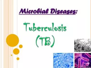
Microbial Diseases of the Lower Respiratory System:Tuberculosis • Mycobacterium tuberculosis • Acid-fast rod • Obligate aerobe • Generation time: > 20 hr • Transmitted from human to human • Airborne droplets reach alveoli • Bacilli are usually phagocytized and killed by macrophages Figure 24.9
Microbial Diseases of the Lower Respiratory System:Tuberculosis Some bacilli may survive inside macrophages More macrophages are recruited… -ineffective at killing -walled-off lesion (tubercle) Figure 24.10.1
Microbial Diseases of the Lower Respiratory System:Tuberculosis Weeks later, many macrophages die -release bacilli into center of tubercle -bacilli do not grow well here -may heal (calcified lesions) -may become dormant infection Figure 24.10.2
Microbial Diseases of the Lower Respiratory System:Tuberculosis Air-filled cavity may form in mature tubercle -active growth of bacilli Cavity grows and may rupture, releasing bacilli into bronchiole -disseminated throughout lungs, blood and lymphatics TB infection in other tissues Figure 24.10.3
Microbial Diseases of the Lower Respiratory System:Tuberculosis • Diagnosis: Tuberculin skin test screening • + = current or previous infection (or vaccination) • Followed by X-ray or CT, acid-fast staining of sputum, culturing bacteria Acid-fast bacillus (AFB) smear (sputum) Figure 24.11 www.cdc.gov
Vaccination recommended only for children at high risk Treatment of TB: Prolonged multiple antibiotic therapy Prolonged naturepatients less likely to complete prescribed regimenemergence of multi-drug resistant TB (MDR-TB) Also XDR-TB (Extensively drug resistant TB) Microbial Diseases of the Lower Respiratory System:Tuberculosis
Microbial Diseases of the Digestive System:Dental Caries, Food poisoning and Helicobacter
>300 species in mouth Large intestine: 100 billion bacteria/gram feces ~40% fecal mass is microbial cell material Assist in polysaccharide breakdown, some synthesize vitamins Mostly anaerobes and facultative anaerobes Bacteriodes, E. coli, Enterobacter, Klebsiella, Proteus Normal Microbiotaof the Digestive System
Bacterial Diseases of the Upper Digestive System:Dental Caries Figure 25.3b
Dental plaque diversity Prescott’s Principles of Microbiology www.gaba.com
Bacterial Diseases of the Upper Digestive System:Dental Caries Figure 25.4
Symptoms usually include diarrhea, gastroenteritis, dysentery Often with abdominal cramps, nausea and vomiting Dysentery: severe diarrhea with blood or mucus Treated with fluid and electrolyte replacement Infection: pathogen enters GI tract and multiplies Incubation from 12 hr to 2 wk Time for colonization, growth and toxin production Typically fever evolves Intoxication: ingestion of preformed toxin Symptoms appear 1-48 hr after ingestion No colonization is necessary Bacterial Diseasesof the Lower Digestive System
Staphylococcal Food Poisoning • Staphylococcus aureus: one of the most common causes • Onset of food poisoning symptoms ~1-6 hours after ingestion of contaminated food • Intoxication • S. aureus is tolerant of high osmotic pressure and low moisture • Somewhat resistant to heat • Most competitors are eliminated (cooking, osmotic pressure) • Multiplies on food, releasing enterotoxin as it grows • Enterotoxin type A: Superantigen exotoxin • Survives up to 30 minutes of boiling! • Triggers vomiting reflex; cramps and diarrhea follow • Recovery within 24 hours
Staphylococcal Food Poisoning S. aureus is present on skin, in nasal secretions Contaminated hands Best prevention strategy: adequate refrigeration Figure 25.6
Sources: contaminated, undercooked meat; raw vegetables Pathogenic E. coli strains: fimbriae for attachment and toxins that cause GI disturbance (gastroenteritis) Low infective dose: <100 bacteria Attach to intestinal mucosa and release toxin into lumen Infection Escherichia coli Gastroenteritis
Enterohemorrhagic E. coli (EHEC): produce Shiga toxin ~50% of feedlot cattle may have enterohemorrhagic strains in their intestines (asymptomatic) E. coli O157:H7 serotype most common cause of outbreaks in US O = cell wall antigen H = flagellar antigen Severe cases (~6%): severe colon inflammation with bleeding (hemorrhagic colitis) Can progress to affect kidneys (hemolytic uremic syndrome) Escherichia coli Gastroenteritis www.sciencenews.org
HelicobacterPeptic ulcer disease • Helicobacter pylori • Cause of majority (70-95%) of peptic ulcer disease cases • Not identified until ~1983 • B. Marshall and Koch’s Postulates • ~40% of adults harbor H. pylori • Only 1-15% develop ulcers • Neutralizes stomach acids so it can thrive (urease) • Causes a drop in protective gastric mucus production Figure 11.11
Helicobacter Peptic ulcer disease Figure 25.13
Helicobacter Peptic ulcer disease H. pylori increases risk of stomach cancers 70-90% of stomach cancers are associated with chronic H. pylori infections www.helico.com
Urinary bladder and upper urinary tract sterile >1,000 bacteria/ml or 100 coliforms/ml of urine indicates urinary tract infection Normal Microbiota of the Urinary System
Urinary Organs Valves to prevent backflow from bladder (shields kidneys from infections) Figure 26.1
Cystitis: infection of the urinary bladder Difficult, painful urination (dysuria) Presence of white blood cells in urine (pyuria) Eight times more common in females vs. males Shorter urethra that’s closer to anal opening Often caused byE. coli Antibiotic-sensitivity tests may be required before treatment 25% untreated cases lead to pyelonephritis Inflammation of one/both kidneys If chronic, scar tissue develops, impairs kidney function 75% due to E. coli Diseases of the Urinary System:Cystitis & Pyelonephritis