Download
1 / 1
10 likes | 15 Views
A. Only one surviving mouse. p<0.0001. p<0.005. n =6 for all groups. B. A. B. mKap. cKap. Association Rate Constant K a (M -1 s -1 ). 3.64x10 4. 3.40x10 4. Dissociation Rate Constant K d (s -1 ). 7.44x10 -4. 7.58x10 -4. K A (M -1 ). 4.87x10 7. 4.48x10 7. Equilibrium

E N D
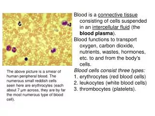
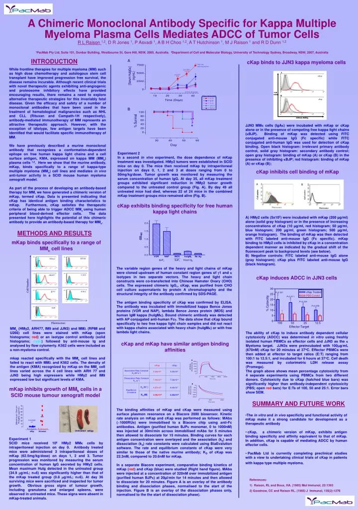
A Only one surviving mouse p<0.0001 p<0.005 n=6 for all groups B A B mKap cKap Association Rate Constant Ka (M-1s-1) 3.64x10⁴ 3.40x10⁴ Dissociation Rate Constant Kd (s-1) 7.44x10⁻⁴ 7.58x10⁻⁴ KA (M-1) 4.87x10⁷ 4.48x10⁷ Equilibrium Constants Untreated mKap KD (M) 2.06x10⁻⁸ 2.23x10⁻⁸ P=0.0065 ** X=0.8mg/mL X=34.9mg/mL INTRODUCTION cKap binds to JJN3 kappa myeloma cells While frontline therapies for multiple myeloma (MM) such as high dose chemotherapy and autologous stem cell transplant have improved progression free survival, the disease remains incurable. Although recent clinical trials with novel therapeutic agents exhibiting anti-angiogenic and proteosome inhibitory effects have provided encouraging results, there remains a need to explore alternative therapeutic strategies for this invariably fatal disease.Given the efficacy and safety of a number of monoclonal antibodies that have been used in the treatment of hematological malignancies such as NHL and CLL (Rituxan and Campath-1H respectively), antibody-mediated immunotherapy of MM represents an attractive therapeutic approach. However, with the exception of idiotype, few antigen targets have been identified that would facilitate specific immunotherapy of MM. We have previously described a murine monoclonal antibody that recognizes a conformation-dependent epitope on free human kappa light chains and a cell surface antigen, KMA, expressed on kappa MM (MM) plasma cells 1,2. Here we show that the murine antibody, mKap, binds specifically to a range of kappa-type multiple myeloma (MM) cell lines and mediates in vivo anti-tumor activity in a SCID mouse human myeloma xenograft model. As part of the process of developing an antibody-based therapy for MM, we have generated a chimeric version of mKap, termed cKap. Data is presented indicating that cKap has identical antigen binding characteristics to mKap. Furthermore, cKap satisfies the therapeutic criteria of being able to trigger ADCC MMusing human peripheral blood-derived effector cells. The data presented here highlights the potential of this chimeric antibody to provide an antibody-based therapy for MM. A Chimeric Monoclonal Antibody Specific for Kappa Multiple Myeloma Plasma Cells Mediates ADCC of Tumor Cells R L Raison 1,2, D R Jones 1, P Asvadi 1, A B H Choo 1,2, A T Hutchinson 1, M J Raison 1 and R D Dunn 1,2 1PacMab Pty Ltd, Suite 101, Dunbar Building, Westbourne St, Gore Hill, NSW, 2065, Australia. 2Department of Cell and Molecular Biology, University of Technology Sydney, Broadway, NSW, 2007, Australia JJN3 MMk cells (IgAk) were incubated with mKap or cKap alone or in the presence of competing free kappa light chains (kBJP). Binding of mKap was detected using FITC conjugated anti-mouse IgG (Fc specific) while FITC conjugated anti-human IgG was used for detection of cKap binding. Open black histogram: irrelevant primary antibody control,solid gray histogram: secondary antibody control; open gray histogram: binding of mKap (A) or cKap (B) in the presence of inhibiting kBJP; red histogram: binding of mKap (A) or cKap (B); Experiment 2 In a second in vivo experiment, the dose dependence of mKap treatment was investigated. HMy2 tumors were established in SCID mice on day 0. The mice then received mKap by intraperitoneal injection on days 0, 1, 2 and 3 at doses ranging from 0 to 50mg/kg/dose. Tumor growth was monitored by measuring the serum concentration of human IgG. At day 35, all mKap treatment groups exhibited significant reduction in HMy2 tumor growth compared to the untreated control group (Fig. A). By day 49 all untreated mice had died, whereas 22 of 24 mice in the combined mKap treatment groups mice remained alive (Fig. B). cKap inhibits cell binding of mKap cKap exhibits binding specificity for free human kappa light chains A) HMy2 cells (5x10⁵) were incubated with mKap (200 µg/ml) alone (solid gray histogram) or in the presence of increasing concentrations of cKap (10 µg/ml, red histogram; 50 µg/ml, blue histogram; 200 µg/ml, green histogram; 500 µg/ml, orange histogram). The binding of mKap was then detected with FITC labeled anti-mouse IgG (Fc specific). mKap binding to HMy2 cells is inhibited by cKap in a concentration dependent manner as indicated by the gradual shift of the fluorescent peak to background levels (see below). B) Negative controls: FITC labeled anti-mouse IgG alone (gray histogram); cKap plus FITC labeled anti-mouse IgG (black histogram). METHODS AND RESULTS mKap binds specifically to a range of MM cell lines The variable region genes of the heavy and light chains of mKap were cloned upstream of human constant region genes of g1 and k isotypes in two separate vectors. The heavy and light chain constructs were co-transfected into Chinese Hamster Ovary (CHO) cells. The expressed chimeric IgG1, cKap, was purified from CHO cell culture supernatants by protein A chromatography and the structural integrity of the antibody confirmed by SDS-PAGE. The antigen binding specificity of cKap was confirmed by ELISA. The antibody was incubated with immobilized kappa Bence Jones proteins (VOR and NAP), lambda Bence Jones protein (MOS) and human IgM kappa (huIgM). Bound chimeric antibody was detected with AP conjugated anti-human Fc. The data show that cKap bound specifically to two free kappa light chain samples and did not react with kappa chains associated with heavy chain (huIgM) or with free lambda light chains. cKap induces ADCC in JJN3 cells MM (HMy2, ARH77, IM9 and JJN3) and MM (RPMI and U266) cell lines were stained with mKap (open histograms; red) or an isotype control antibody (solid histograms; cyan) followed by anti-mouse Ig and analysed by flow cytomerty. K562 cells were included as a non-myeloma control. mkap reacted specifically with the MM cell lines and failed to react with MM and K562 cells. The density of the antigen (KMA) recognized by mKap on the MM cell lines varied across the 4 cell lines with ARH 77 and JJN3 being high expressers while HMy2 and IM9 expressed low but significant levels of KMA. The ability of cKap to induce antibody dependent cellular cytotoxicity (ADCC) was determined in vitro using freshly isolated human PBMCs as effector cells and JJN3 as the k Myeloma target. JJN3s were preincubated with 100mg/mL (670nM) cKap for 20 minutes at 37oC. Effector PBMCs were then added at effector to target ratios (E:T) ranging from 100:1 to 12.5:1, and incubated for 6 hours at 37oC. Cell death was measured by colorimetric LDH release assay (Promega). The graph above shows mean percentage cytotoxicity from 4 separate experiments using PBMCs from two different donors. Cytotoxicity due to cKap (hatched blue bars) was significantly higher than antibody-independent cytotoxicity (PBS; open red bars) for E:Ts of 100, 50 and 25:1. Error bars show SEM. cKap and mKap have similar antigen binding affinities mKap inhibits growth of MMk cells in a SCID mouse tumour xenograft model SUMMARY AND FUTURE WORK The binding affinities of mKap and cKap were measured using surface plasmon resonance on a Biacore 2000 biosensor. Kinetic rate analysis on mKap and cKap was performed as follows: MAbs (~1000RUs) were immobilised to a Biacore chip using anti-Fc antibodies. Antigen (purified human BJPk monomer, 0 to 1000nM) was injected at 30ml/minacross immobilised mAbs for 5 minutes then allowed to dissociate for 15 minutes. Binding curves for each antigen concentration were overlayed and the association (ka) and dissociation (kd) rate constants were calculated using BiaEvalution software. The rate and equilibrium constants of cKap were very similar to those of the native murine antibody; KD of cKap was 22.3nM, compared to 20.6nM for mKap. In a separate Biacore experiment, comparative binding kinetics of mKap (red) and cKap (blue) were studied (Right hand figure). MAbs were injected at a concentration of 320nM over immobilized antigen (purified human BJPk) at 20µl/min for 14 minutes and then allowed to dissociate for 20 minutes. Figure A is an overlay of the antibody binding and dissociation phases, normalised to the start of the injection. Figure B is an overlay of the dissociation phases only, normalised to the the start of dissociation phase). • The in vitro and in vivo specificity and functional activity of mKap make it a strong candidate for development as a therapeutic antibody • cKap, a chimeric version of mKap, exhibits antigen binding specificity and affinity equivalent to that of mKap. In addition, cKap is capable of mediating ADCC by human effector cells. • PacMab Ltd is currently completing preclinical studies with a view to undertaking clinical trials of cKap in patients with kappa type multiple myeloma. Experiment 1 SCID mice received 106 HMy2 MM cells by intraperitoneal injection on day 0. Antibody treated mice were administered 3 intraperitoneal doses of mKap (62.5mg/kg/dose) on days 1, 2 and 3. Tumor progression was monitored by measuring the serum concentration of human IgG secreted by HMy2 cells. Mean maximum HuIg detected in the untreated group (34.9 g/mL; n=6) was significantly higher than that of the mKap treated group (0.8 g/mL; n=6). At day 36 surviving mice were sacrificed and inspected for tumor growth. Obvious gross signs of tumour growth, including granuloma and enlarged spleens, were observed in untreated mice. These signs were absent in mKap-treated animals. • References: • Raison, RL and Boux, HA. (1985) Mol Immunol, 22:1393 • 2) Goodnow, CC and Raison RL. (1985) J Immunol, 135(2):1276