Download
1 / 30
330 likes | 561 Views
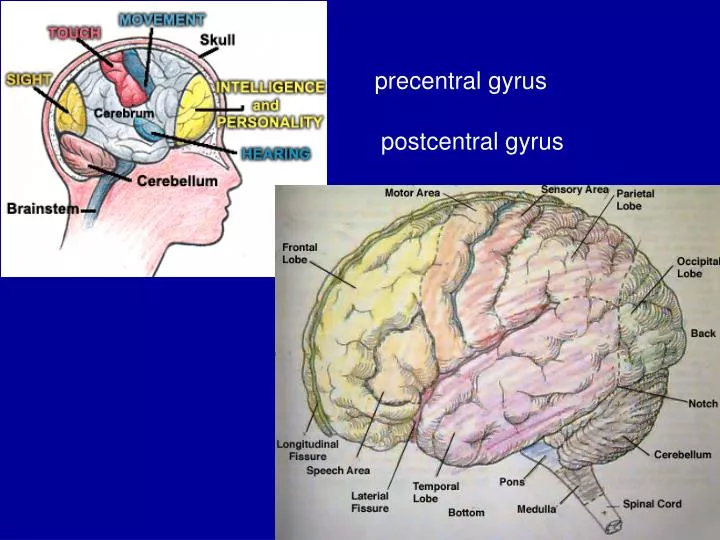
precentral gyrus postcentral gyrus. Main Contents. The thoracic wall Breast Intercostal spaces Mediastinum Concept, boundaries, division Superior mediastinum Posterior mediastinum. Introduction. 1 formation 2 division : Thoracic wall—

E N D
Main Contents The thoracic wall • Breast • Intercostal spaces Mediastinum • Concept, boundaries, division • Superior mediastinum • Posterior mediastinum
Introduction 1 formation 2 division: • Thoracic wall— Anterior region of thorax, anterolateral region of thorax and posterior region of thorax. • Thoracic cavity—mediastinum, left and right lung and pleura cavity.
The thoracic wall superficial structures 1)skin 2)superficial fascia 3)cutaneous nerve : • The supraclavicular nerve • Anterior cutaneous branches of intercostal nerve • Lateral cutaneous branches of intercostal nerve
superficial structures 4) superficial vessels • artery: • perforating branches of internal thoracic artery • the branches of posterior intercostal A. • Vein: • → internal thoracic V. and posterior intercostal V. • The V. of thoracic and abdominal wall→lateral thoracic V.
Deep structures Intercostal spaces • Position • Contents: 1) The intercostal externus 2) The intercostal internus 3) The intercostal intimus 4) The intercostal nerves:
Intercostal spaces 5) Veins and Arteries 6)The relationship of the intercostal nerve and vessels in the costal groove: — V. A. N..
Mediastinum Boundaries • Anterior: the sternum and costal cartilages. • Posterior: the thoracic vertebrae • Two sides: the mediastinal pleurae. • Superior: the thoracic inlet. • Inferior: the diaphragm
Mediastinum 2 Division • four parts of the mediastinum
superior mediastinumglandular plane venous plane arterial-nervous plane visceral plane lymphatic plane • anterior layer • thymus or its rudiment; • right and left brachiocephalic V.; • superior vena cava; • middle layer(arterial layer) • aorta and its three branches; • right and left phrenic and vagus nerves; • posterior layer • trachea; • esophagus; • Left recurrent laryngeal nerve • thoracic duct.
1) Thymus Relationship ―two side: ―upper boundary: ―lower boundary: External feature ―two unequal lateral lobes and a pyramidal lob. 2) Superior vena cava 3) The aortic arch and its branches superior mediastinum
superior mediastinum 4)The thoracic part of trachea in front of the the thoracic part of trachea : ① the sternal manubrium; ② origins of sternohyoid and sternothyroid muscles; ③ thymus; ④ left brachiocephalic V., ⑤ aortic arch; ⑥ brachiocephalic trunk; ⑦ left common carotid A. ⑧ cardiac plexus and lymph nodes.
Main organs: The Posterior Mediastinum ① principal bronchi; ② thoracic part of esophagus; ③ thoracic aorta; ④ thoracic duct; ⑤ azygos and hemiazygos V.; ⑥ vagus nerves; ⑦ sympathetic trunk and lymph nodes.
The Posterior Mediastinum The thoracic part of esophagus Relation • Anteriorly: • Posteiorly: • Left side: • Right side:.
The thoracic part of esophagus 2) Blood supply Arteries: • Superior part • Inferior part Veins: →azygos vein, hemiazygos vein,accessory hemiazygos vein → Superior vena cava
The thoracic part of esophagus 3) Lymphatic drainage • Superior part • Middle part • Inferior part • Part of it 4) Nerves
The Sympathetic Trunk • 10-20 ganglia and interganglionic fibers. • upper 4th-5th ganglions→esophageal, cardic, pulmonary plexuses. • 5th-9th ganglions→greater splanchnic N. →celial ganglion • 10th-12th ganglions→lesser splanchnic N. →aorticorenal ganglion
Thoracic aorta 1)position and passage 2)branches Parietal branches and visceral branches 3)relation anteriorly: posteriorly: right: left:
Question ? What are they in the posterior mediastinum?
Summary The thoracic wall • Breast • Intercostal spaces Mediastinum • Superior mediastinum • Posterior mediastinum