Download
1 / 39
390 likes | 502 Views
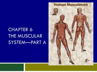
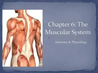
Chapter 6 The Muscular System. Biology 112 Tri-County Technical College Pendleton, SC. Functions of Muscles. Essential function of muscle is CONTRACTION (shortening) Separates it from other body tissue Responsible for essentially all body movement Can be viewed as “machines” of body

E N D
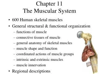
Chapter 6 The Muscular System Biology 112 Tri-County Technical College Pendleton, SC
Functions of Muscles • Essential function of muscle is CONTRACTION (shortening) • Separates it from other body tissue • Responsible for essentially all body movement • Can be viewed as “machines” of body • Makes up nearly half of the body’s mass
Types of Muscle • Skeletal, Cardiac, and Smooth • Share certain characteristics • Muscle cells are elongated and are called MUSCLE FIBERS • Ability to shorten depends on TWO types of MYOFILAMENTS • Muscle cells =“microfilaments of cytoskeleton” • “Myo,” “Mys,” and “Sarco” refer to muscle • Sarcoplasm-cytoplasm of muscle cells
Skeletal Muscle • Skeletal muscle fibers packed into organs called skeletal muscles that attach to skeleton • Known as striated muscle = fibers appear striped • Only muscle subject to CONSCIOUS control • Each fiber enclosed in CT sheath called ENDOMYSIUM • Several sheathed muscle fibers wrapped by membrane called PERIMYSIUM to form bundle of fibers called a FASCICLE
Skeletal Muscle, cont. • Many fascicles bound by EPIMYSIUM which covers entire muscle • EPIMYSIS blend into strong, cordlike TENDONS (sheetlike aponeuroses) which attach muscles indirectly to bone, cartilages, or connective tissue covering of each other • Spend some time on 6.1; page 164
Sarcomere & Myofibrils • Plasma membrane of muscle cell called SARCOLEMMA • Long ribbonlike organelles called MYOFIBRILS nearly fill cytoplasm • Alternating light (I) and dark (A) bands along length of myofibril = striped appearance • Light I band has midline interruption; a darker area called the Z DISC (line) • Dark A band has lighter central area called the H zone
Sarcomere, cont. • Myofibrils are chains of tiny contractile units called SARCOMERES which are aligned end to end like boxcars in train • IT is arrangement of smaller MYOFILAMENTS within sarcomere that actually produces banding pattern • Spend some quality time on Figure 6.3; page 167
Myofilament Arrangement • Each sarcomere contains two types of myofilaments • Large thick filaments (MYOSIN) extend entire length of dark A band • Midparts of thick filaments are smooth but ends are studded with small projections (myosin heads or cross bridges) • Thin filaments (ACTIN) anchored to Z line which is actually disclike membrane • Light I band is area that includes parts of two adjacent sarcomeres and contains ONLY thin filaments
Myofilament Arrangement, cont. • Thin filaments overlap ends of thick filaments but DO NOT extend into middle of relaxed sarcomere • Thus central region (H zone) looks bit lighter • Contraction occurs, actin containing filaments slide toward each other into center of sarcomere and light zones disappear • Actin and myosin filaments completely overlap
Sarcomere Contraction • Fibers activated by NS, cross bridges on myosin attach to myosin binding sites of actin filaments • Attaches and detaches several times during contraction and PULLS thin filaments toward center of sarcomere • Event occurs simultaneously in sarcomeres throughout cell, muscle cell shortens • Z lines move closer together • H zone disappears • A bands move closer together but do NOT change in length • Millions of sarcomeres in millions of fibers = contraction of entire skeletal muscle
Connective Tissue Wrappings • Skeletal muscle wrapped by connective tissue • ENDOMYSIUM wraps each individual muscle fiber (cell) • PERIMYSIUM wraps bundles of fibers into a FASCICLE • EPIMYSIUM covers the entire muscle • Epimysium is continuous with tendons or aponeuroses
Smooth Muscle • Has NO striations and is involuntary • Walls of hollow visceral organs like stomach, urinary bladder, digestive tract, bronchi, uterus, and blood vessels • Visceral-nonstriated-involuntary • Spindled-shaped fibers with single nucleus and arranged in sheets/layers • Contractions SLOW and SUSTAINED-does Not tire easily • Movement of food through digestive tract, emptying bowels and bladder, & maintenance of blood pressure
In a heartbeat…so to speak • Cardiac muscle best described as CARDIAC, STRIATED, AND INVOUNTARY • Branching cells joined by special junctions called intercalated disks • Cardiac muscle arranged in spiral shape • Allows contractions to be closely coordianted
Nerve Aspects • Each muscle fiber must be stimulated separately by nerve impulses to contract • MOTOR UNIT-one motor neuron (nerve cell) and all the skeletal muscle cells it stimulates • Threadlike extensions of neuron (axon/nerve fiber) branch into number of axonal terminals at muscle • Each axonal terminal forms junctions with sarcolema of different muscle cell • Junctions called neuromuscular junctions
Nerve Aspects, cont. • Nerve endings and muscle cells’ membranes NEVER TOUCH • Gap between them called synaptic cleft and is filled with interstitial fluid • Nerve impulse reaches axonal terminals, a neurotransmitter is released • Specific neurotransmitter that simulates muscle cells is ACETYLCHOLINE (Ach) • Acetylcholine diffuses across synaptic cleft
Nerve Aspects, cont. • Acetylcholine attaches to receptors on sarcolemma • If enough released, sarcolemma becomes temporary permeable to sodium ions (Na+) which rush into muscle cell • Generates electrical current called action potential • AP travels over entire surface of sarcolemma conducting impulse from one end of cell to the other • Result is CONTRACTION of the cell
Sliding Filament Theory • Nerve impulseneuromuscular junction acetylcholine releasedAP in sarcolemma • AP in sarcolemma causes sarcoplasmic reticulum to releases stored calcium ions into sarcoplasm • Calcium ions cause cross-bridges to from • Thin myofilaments (actin) pulled over thick (myosin) myofilaments • Energy provided by ATP
SF Theory, cont. • Sacromere contracts • AP ends, calcium ions reabsorbed • Cross-bridges turn loose & sarcomere relaxes • Neurotransmitter acetylcholine degraded by enzymes in synaptic cleft • Prevents continuous stimulation of muscle fiber • Acetylcholinesterase (care for some Raid, anyone?)
To twitch or not to twitch • Muscle fiber contracts in all-or-none fashion • Whole muscles do NOT contract that way • Skeletal muscles are organs composed of 1000s of muscle cells which react to stimuli with GRADED RESPONSES (different degrees of shortening) • Graded muscle contractions produced in 2 ways • Changing speed of muscle stimulation • Changing number of muscle cells being stimulated
Twitching time, cont. • Muscle twitch is single, brief, jerky contraction that occurs as result of certain nervous system problems • NOT the way muscle normally operates • Single stimulus-contraction-relaxation sequence in muscle fiber • DOES NOT accomplish anything useful in skeletal muscle
Twitch and more, cont. • Incomplete tetanus results when nerve impulses delivered to muscle at very high rate • Delivered so rapidly cells do not get chance to relax completely between stimuli • Stimulation continues and muscle never allowed to relax completely will cause tension to peak • Muscle producing peak tension during rapid cycles of contraction/relaxation said to be in incomplete tetanus
Enough on twitching already • Complete tetanus results when muscle is stimulated so rapidly that NO evidence of relaxation is seen • Contractions are completely smooth and sustained • Complete tetanus major role = smooth and prolonged muscle contractions • Force of muscle contraction depends on how many of its cells are stimulated • Few cells = contraction as whole is slight • All cells = muscle contraction as strong as it can be
Fatigue and Debt • Muscle subject to continual contraction for long time = muscle fatigue occurs • Muscle is fatigued when unable to contract even though still be stimulated • Without rest, active/working muscle begins to tire and contracts more weakly until finally ceases reacting and stops contracting • MF believed to result from oxygen debt that occurs during prolonged muscle activity
Fatigue & Debt, cont. • Work muscle can do and how long it can work w/o becoming fatigued depend on how good its blood supply is • If muscle runs out of O2, it must depend on glycolysis for ATP and converts pyruvic acid to lactic acid • Lack of adequate ATP and >acidity cause muscle to contract less effectively and finally to stop contracting all together
Isotonic Contractions • Isotonic (same tone/tension) contractions most familiar • Myofilaments are successful in sliding movements, muscle shortens, and movement occurs • Bending knee, rotating arms, and smiling are examples of isotonic contractions
Isometric Contractions • Isometric (same measurement/length) contractions are contractions in which muscles do NOT shorten • Mysoin myofilaments skidding their “wheels” and tension in muscle keeps increasing • Trying to slide but muscle pitted against some more/less immovable object(s) • Trying to lift 400 lb dresser along or pushing against immovable wall
Muscle tone • When muscle voluntarily relaxed, some of its fibers are contracting (one group then another) • As result, muscle remains firm, healthy, and ready for action • This state of continuous partial contractions is called MUSCLE TONE • Is the result of different motor units scattered through muscle being stimulated by nervous system in systematic way
Muscle tone, cont. • If nerve supply to muscle is destroyed, muscle NO longer stimulated in this manner • It loses TONE, and becomes paralyzed • Soon after, becomes flaccid (soft/flabby) and begins to atrophy (waste away)