Download
1 / 20
210 likes | 519 Views
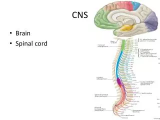
Diseases of CNS. Topics:. H ydrocephalus. Cerebrovascular diseases. Brain abscess. Neurodegenerative diseases. Brain tumors. Hydrocephalus. ILOs. Understanding definition of hydrocephalus. Discussing causes of hydrocephalus. Describing morphological features of hydrocephalus.

E N D
Topics: • Hydrocephalus. • Cerebrovascular diseases. • Brain abscess. • Neurodegenerative diseases. • Brain tumors.
ILOs • Understanding definition of hydrocephalus. • Discussing causes of hydrocephalus. • Describing morphological features of hydrocephalus. • Being aware of the complications of hydrocephalus.
* Def: Dilation of ventricular system due to excess C.S.F volume associated with atrophy of the brain tissue.. * Causes: I. Increased CSF production: choroid plexus papilloma or choroiditis. II. Obstruction of CSF flow: a. Congenital causes: 1. Narrowing of foramina of the ventricles or aqueduct of sylvius.
2. Arnold - Chiari malformation: down-word displacement of medulla and cerebellum into foramen magnum with block of the foramina of fourth ventricle. b. Acquired causes: due to post-meningitis fibrosis or space occupying lesions. III. Defective absorption through arachnoid villi: • Congenital causes: agenesis, aplasia or hypoplasia of arachnoid villi. • Acquired causes: arachnoid villi fibrosis – superior sagital sinus thrombosis.
* Types: 1. Communicating Hydrocephalus: When obstruction occur outside the ventricular system. 2. Non communicating Hydrocephalus: When obstruction occurs within the ventricles. 3. Hydrocephalus Ex vacuo: Refers to dilatation of ventricular system with compensatory increase in CSF volume secondary to brain atrophy.
* Morphology: • Dilated ventricles. • Increased C.S.F pressure leads to peri-ventricular interstitial edema. • Pressure atrophy of the brain tissue. • Skull changes: A. Children: • Thin bone. • Separated sutures. • Enlarged skull. • Mental deficiency. B. Adult: • The inner vault of the skull shows varying degrees of convolutional markings (mouse eaten appearance).
* Signs and Symptoms: • Symptoms of increased intracranial pressure: include: • Headache. • Vomiting. • Nausea. • Papilledema. • Sleepiness or coma. • Cerebellar tonsill herniation, with resulting life threatening brain stem compression.
Early symptoms may also include: • Eyes that appear to gaze downward (Sundowning) • Irritability • Seizures • Separated sutures • Sleepiness • Vomiting
Symptoms that may occur in older children can include: • Changes in personality, memory, or the ability to think • Changes in facial appearance and eye spacing • Uncontrolled eye movements • Difficulty feeding • Excessive sleepiness • Headache • Irritability • Loss of bladder control (urinary incontinence) • Loss of coordination and trouble walking • Muscle spasticity (spasm) • Slow growth • Slow or restricted movement • Vomiting
* Treatment: - Hydrocephalus treatment is surgical. • Cerebral shunts: It involves the placement of a ventricular catheter, into the cerebral ventricles to bypass the flow obstruction/malfunctioning arachnoidal granulations and drain the excess fluid into other body cavities, from where it can be resorbed.
Most shunts drain the fluid into the peritoneal cavity (ventriculo-peritoneal shunt), but alternative sites include the right atrium (ventriculo-atrial shunt), pleural cavity (ventriculo-pleural shunt), and gallbladder. A shunt system can also be placed in the lumbar space of the spine and have the CSF redirected to the peritoneal cavity (Lumbar-peritoneal shunt).
* Shunt complications: • Shunt infection. • Shunt malfunction. • Shunt failure.
Other treatments may include: • Antibiotics are given if there are signs of infection. Severe infections may require the shunt to be removed.
Follow up: The child will need regular check-ups to make sure there are no further problems. Tests are regularly done to check the child's developmental and for intellectual, neurological, or physical problems.