Download

1 / 15
260 likes | 1.49k Views
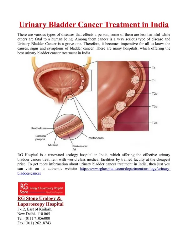
Development of the urinary bladder and urethra . Dr. Sanaa Alsharawi / Dr. Essam Salama . Objectives . At the end of the lecture the student is able to; Describe the cloaca and the formation of the urogenital sinus.

E N D
Development of the urinary bladder and urethra Dr. Sanaa Alsharawi / Dr. Essam Salama
Objectives • At the end of the lecture the student is able to; • Describe the cloaca and the formation of the urogenital sinus. • Discuss the division of the urogenital sinus into various parts and name the adult organs that are derived from each part. • Describe how the caudal parts of the mesonephric ducts and ureters are absorbed into the urogenital sinus and the significance of this embryonic event. • Discuss the position of the urachus and its significance and fate. • Describe the various anomalies concerned with the urinary bladder and urethra.
Cloaca The cloaca is the dilated terminal part of the hind gut. It receives the allantoisand the mesonephric ducts. Its floor is closed by the cloacal membrane.
Cloaca A mesodermal urorectal septum divides the cloaca and the cloacal membrane into: Ventral part; the primitive urogenital sinus that communicates with the allantois and the mesonephric ducts. Its floor is the urogenital membrane. Dorsal part; forms the rectum and upper part of anal canal. Its floor is the anal membrane.
Primitive urogenital sinus Is divided into three parts; A cranial;vesical part; forms most of the bladder and continuous with the allantois. A middle;pelvic part; forms main part of male urethraand entire female urethra. A caudal;phallic part grows towards genital tubercle.
Urinary bladder It develops mainly from the vesical part of the urogenital sinus. The trigone is derived from the absorbed caudal ends of the mesonephric ducts. The epithelium is endodermal in origin. The other layers are derived from the splanchinic mesoderm.
Urinary bladder The allantois is at first continues with the bladder , then it becomes a thick fibrous cord urachus which extends from apex of the bladder to the umbilicus, in adult it is represented by the median umblical ligament. After absorption of the mesonephric ducts to form the trigone, the ureters open separately in the bladder. In infants and children the bladder is an abdominal organ , it begins to enter the greater pelvis at about 6 years and becomes a pelvic organ until after puberty.
Urethra Indifferent stage ; The genital tubercle (mesenchymal elevation) develops at the cranial end of the cloacal membrane. Two urethral folds, develop on either side of the urogenital membrane. Laterally two labioscrotal folds develop on either side of the urethral folds. 2 urethral folds in male fuse with each other to close the penile urethra. 2 urethral folds in female remain separate to form labia minora. (Labioscrotal folds) Penile Urethra
Female Urethra The entire female urethra is derived from endoderm of the pelvicpart of the urogenital sinus. The external urethral orifice opens dorsal to the glans clitoris.
Male Urethra The genital tubercle elongates forming the phallus, which is the precursor of the penis. Most of the male urethra : prostatic, membranous and spongy parts is derived from endoderm of the pelvic middle part of urogenitalsinus. The distal part of male urethra in glans penis starts as ectodermalsolid cord that grows towards the root of penis to meet the spongy urethra , later it canalizes.
Anomalies • Urachal anomalies. • Urethral anomalies. • Extrophy of the bladder (Ectopiaevesica); exposure of the posterior wall of the bladder due to a defect in the anterior abdominal wall and anterior wall of the bladder.
Urachal anomalies A, Urachal cystpersistence or remnant of epithelial lining of urachus. B, Urachal sinus,discharge serous fluid from the umblicus. C, Urachal fistula,the entire urachus remains patent and allows urine to escape from the umbilicus.
Urethral Anomalies 1-Hypospadius : is the most common anomaly, with incomplete fusion of the urethral folds, and abnormal openings of the urethra occur along the ventral (inferior) aspect of the penis. 2-Epispadius : is a rare abnormality, in which the urethral meatus is found on the dorsum of penis, it is most often associated with extrophy of the bladder.
1. The urinary bladder is mainly developed from : a. Vesical part of the urogenital sinus. b. Pelvic part of the urogenital sinus. c. Pallic part of the urogenital sinus. d. Allantois. 2. Which one of the following forms the entire female urethra ? a. Genital tubercle. b. Allantois. c. Vesical part of the urogenital sinus. d. Pelvic part of the urogenital sinus. 4. The trigone of the urinary bladder is developed from : a. Paramesonephric ducts. b.Mesonephric ducts. c. Allantois. d. Urogenital sinus. 5. The urethra in glans penis is developed from : a. The vesical part of urogenital sinus. b. The pelvic part of urogenital sinus. c. The ectoderm. d. The splanchnic mesoderm.