Download
1 / 89
950 likes | 1.61k Views
Types of hypoxia and management . Dr . Gaurav Dhakate. University College of Medical Sciences & GTB Hospital, Delhi. types. CPR. Surgical consequences. Consequences . OXYGEN DELIVERY TO CELLS. Normal 1000 mls /minute (550 mls /min/m2) of oxygen is transported

E N D
Types of hypoxia and management Dr. GauravDhakate University College of Medical Sciences & GTB Hospital, Delhi
OXYGEN DELIVERY TO CELLS • Normal 1000 mls/minute (550 mls/min/m2) of oxygen is transported Satisfactory delivery to tissues depends on a number of factors: • Adequate alveolar ventilation • Diffusion • macro and micro circulation
ANAESTHETIC FACTORS • Gas exchange abnormalities in the post-operative period occur early or late. • Early post-operative hypoxaemia • alveolar hypoventilation (above), • Ventilation/perfusion mismatching, • Decreased cardiac output and • Increased oxygen consumption due to shivering (induced by volatile agents) • recovery from intra-operative hypothermia. • ‘diffusion hypoxia’
The later onset • functional residual capacity (FRC) • patient’s inability to inspire deeply or cause the patient to be immobilised in bed.eg pain
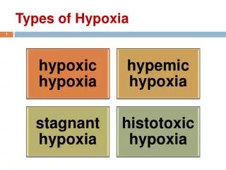
I. HYPOXEMIC (DECREASED TISSUE OXYGEN TENSION) I A. HYPOXEMIC HYPOXIA (INADEQUATE ARTERIAL OXYGEN TENSION AND SATURATION) CAUSES: A. V/Q MISMATCH (EX: COPD, PE) B. SHUNT (EX: ATELECTASIS, PULM. EDEMA) C. HYPOVENTILATION (EX: DRUG INDUCED)
PAo2 (is a result of dynamic equilibrium btw delivery and extraction) BP(high altitude) Minute ventilation(drug overdose) O2 delivery
O2 extraction Mixed venous o2 content and Pvo2 Pulmonary capillary blood flow= CO
Mixed venous Po2 [Pvo2] Pvo2 o2 extraction Pao2 hypoxia Less volume of blood presented to tissues per unit time so more o2 will be extracted by tissues
Management FiO2 Barometric pressure Maintain MV Optimize CO Adjunctive Improve lung cond.
Signs of resp. fatigue , circulatory collapse Prob.= PAO2 Aim= PAO2 Intubate+mech vent.(PEEP) ECMO
V/Q mismatch Diffusion capacity Supplement O2 CPAP by face mask Tracheal intubation Adjunctive Th.
B. ANEMIC HYPOXIA(DEFICIENT OXYGEN-CARRYING CAPACITY OF THE BLOOD) CAUSES: A. ANEMIA (DECREASED HEMOGLOBIN) B. CARBON MONOXIDE POISONING C. SULFHEMOGLOBIN AND METHEMOGLOBIN
At normal Hb conc. ,20 ml of o2 is carried by 1 dl(100 ml) of blood. • At tissue site,o2 consumption is same and perfusion is also same ,but due to decrease in o2 content,low Po2 in capillary adjacent to the tissues • Decrease pressure head for diffusion of o2 to tissues • Tissue hypoxia
CONTENT VS TENSION (PaO2) • A. CONTENT= TOTAL AMOUNT OF OXYGEN CARRIED IN BLOOD NORMAL = 20.7 VOL% CALCULATION: CaO2 = [%sat x l.39 x hb] + [PaO2 x 0.003] EXAMPLES/NORMAL NORMALHb% = 15 GM%, 0.98 02 SAT = PaO2 = 100mmHg [1.39 X 0.98 x 15] + [100 x 0.003] = 20.7 mg/dl ANEMIAHb%, %sat = 98%, PaO2 = 100mmHg [1.39 x 0.98 x 10] + [100 x 0.003] = 14.2 mg/dl HYPOXEMIAHb% =15 gm%, %Sat=85%, PaO2=50mmHg [1.39 x0.85 x 15] = [50 x 0.003] = 18.0mg/dl NORMAL MIXED VENOUS CONTENT = 15% ARTERIAL VENOUS DIFFERENCE (A-V) = 5 VOL%
Carboxyhaemoglobin Causes: Smoking. Auto exhaust,fire
Methemoglobin Inherited MOA: same as carboxyhb
MOA:normalhb with a sulphur atom incorporated into porphyrin ring
CONTINUED C. CIRCULATORY HYPOXIA (DECREASE PERIPHERAL CAPILLARY BLOOD FLOW) CAUSES : A. DECREASED CARDIAC OUTPUT B. VASCULAR INSUFFICIENCY (SEPSIS) D. HISTOTOXIC HYPOXIA (DECREASED UTILIZATION OF OXYGEN AT THE CELL LEVEL) CAUSES: A. CYANIDE POISONING B. ALCOHOL POISONING (RARE)