Download
1 / 35
350 likes | 471 Views
The Autonomic Nervous System. Chapter 17. Introduction. Makes all routine adjustments in physiological systems. Consists of visceral motor (efferent) neurons The ANS pathway from the CNS to the effector always involves 2 neurons synapsing in an autonomic ganglion

E N D
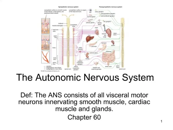
The Autonomic Nervous System Chapter 17
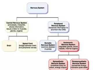
Introduction • Makes all routine adjustments in physiological systems. • Consists of visceral motor (efferent) neurons • The ANS pathway from the CNS to the effector always involves 2 neurons synapsing in an autonomic ganglion • Preganglionic (neuron #1) – cell body is in the CNS, axon extends to the ganglion outside the CNS • Postganglionic (neuron #2) – cell body is in the ganglion, axon extends to the visceral effector
Nerve Fibers of the ANS • Preganglionic (neuron #1) • Always myelinated • Neurotransmitter is always ACh • Postganglionic (neuron #2) • Always nonmyelinated • Neurotransmitter is Ach or norepinephrine
Subdivisions of the ANS • Sympathetic division (thoracolumbar) • Cell bodies for all the neurons #1 reside in the thoracic and lumbar portions of the spinal cord. • T1 – L2 • Stimulates heart beat & tissue metabolism, increases alertness, prepares the body to deal with emergencies (“fight or flight” division)
Subdivisions of the ANS • Parasympathetic division (craniosacral) • Cell bodies reside in the brain stem (cranial nerves) or in the sacral portion of the spinal cord. • Slows the heart rate, inhibits senses, prepares the body for rest and relaxation; (“rest and digest” division).
Sympathetic Chain Ganglia • A chain of ganglia that run alongside the spinal cord • Extends on both sides of the vertebral column • Carries preganglionic fibers and cell bodies of postganglionic neurons
Anatomy of the Sympathetic Chain • Rami communicates from the spinal nerves connect to the chain • Other nerves (splanchnic) project from the chain
Routes of Preganglionic Axons • Cell bodies of neurons #1 lie in the lateral gray horns of the spinal cord • The axons of neurons #1 leave the spinal cord via the ventral root • These axons pass to the spinal nerve • Axons leave the spinal nerve via the white and gray branches (rami communicates) • Connect with the sympathetic chain ganglia
Routes of Preganglionic Axons • There are 3 possible routes that sympathetic neurons #1 may follow • Possibility #1: synapses with the ganglion at that level • Neuron #2 leaves at that level via the gray ramus communicans, rejoins the same level spinal nerve
Routes of Preganglionic Axons • Possibility #2: neuron #1 goes up or down the chain and synapses at some other level. • Neuron #2 leaves at that level via the gray ramus communicans, rejoins the spinal nerve at that level. • Possibility #3: neuron #1 does not synapse in the chain but exits by a splanchnic nerve and synapses in a collateral ganglion. • Neuron #2 travels from that ganglion to its destination.
Collateral Ganglia • Location – anterior to the aorta in the abdominopelvic cavity • Celiac ganglion • Innervates upper abdominal viscera • Superior mesenteric • Innervates middle abdominal viscera • Inferior mesenteric • Innervates lower abdominal & pelvic organs
The Adrenal Medulla • Only preganglionic neurons are in this pathway • Neuron #1 stimulates the medulla, • The medulla releases norepinephrine and epinephrine (adrenaline) to blood
Effects of Sympathetic Stimulation • Widespread • The sympathetic chain allows one preganglionic fiber to synapse with many postganglionic neurons • Enhanced & prolonged by the adrenal medulla
Neurotransmitters • Preganglionic fibers release acetylcholine (Ach) • Cholinergic • Postganglionic fibers (most) release norepinephrine (NE) • Adrenergic • Adrenal medulla releases norepinephrine and epinephrine (adrenaline)
Membrane Receptors & Sympathetic Function • 2 types of receptors in synapses • The same neurotransmitter can have different effects • Alpha receptors cause a rise in intracellular calcium • Respond more than beta to NE • Beta receptors cause changes in the metabolic activity of the target cells
Summary of Sympathetic Division • Cell bodies are found in the thoracic and lumbar portions of the spinal cord • Preganglionic fibers are short, connect to the sympathetic chain, and synapse with long postganglionic fibers • Preganglionic fibers produce ACh, postganglionic fibers produce NE or Ach • “Fight or flight” division
Organization of the PNS • Cell bodies are in the brain or in the gray matter of the spinal cord (sacral region) • Neurons #1 exit the cranial region through cranial nerves 3, 7, 9, & 10 • Neurons #1 exit the spinal cord through the sacral spinal nerves
Organization of the PNS • Neurons #1 are long and synapse with neurons #2 (short) in ganglia • Ganglia are found on near the visceral effector
Anatomy of the PNS • The cranial nerve fibers involved are motor - control smooth muscle & glands in the upper body • Cranial nerve #3 – lens & pupil • Cranial nerve #7 – lacrimal glands, submandibular & submaxillary glands (salivary) • Cranial nerve #9 – parotid gland (salivary) • Cranial nerve #10 - viscera of thorax & abdomen • Sacral nerves innervate the kidneys, colon, & sex organs
General Functions of the PNS • Prepares the individual for rest and relaxation • “Rest & digest” division • Effects on various organs: • Decreases heart rate • Constricts bronchioles • Increases salivation • Increases motility of stomach • Increases motility of colon • Constricts pupils
Neurotransmitter • Both preganglionic and postganglionic fibers release acetylcholine • Causes localized and short-term effects
Summary of the Parasympathetic Division • Cell bodies are found in the brain and in the sacral region of the spinal cord • Preganglionic fibers are long and synapse with short postganglionic fibers on or near the target viscera • Both preganglionic and postganglionic fibers produce Ach • “Rest & digest” division
Relationship Between the Sympathetic and Parasympathetic Divisions • Most organs receive dual innervation • Visceral organs are intrinsically excited • ANS either increase excitation or inhibit the activity • Eg. Sympathetic fibers increase heart rate, parasympathetic fibers decrease heart rate