Download
1 / 17
300 likes | 1.11k Views
Basic Physics of Ultrasound. WHAT IS ULTRASOUND?. Ultrasound or ultrasonography is a medical imaging technique that uses high frequency sound waves and their echoes. Known as a ‘pulse echo technique’

E N D
WHAT IS ULTRASOUND? • Ultrasound or ultrasonography is a medical imaging technique that uses high frequency sound waves and their echoes. • Known as a ‘pulse echo technique’ • The technique is similar to the echolocation used by bats, whales and dolphins, as well as SONAR used by submarines etc.
In ultrasound, the following events happen: • The ultrasound machine transmits high-frequency (1 to 12 megahertz) sound pulses into the body using a probe. • The sound waves travel into the body and hit a boundary between tissues (e.g. between fluid and soft tissue, soft tissue and bone). 3. Some of the sound waves reflect back to the probe, while some travel on further until they reach another boundary and then reflect back to the probe . 4. The reflected waves are detected by the probe and relayed to the machine.
The machine calculates the distance from the probe to the tissue or organ (boundaries) using the speed of sound in tissue (1540 m/s) and the time of the each echo's return (usually on the order of millionths of a second). 6. The machine displays the distances and intensities of the echoes on the screen, forming a two dimensional image.
So…. • All the energy comes from the transducer • All we “see” are reflections and scatter.
SOUND Sound waves consist of mechanical vibrations containing condensations (compressions) & rarefactions (decompressions)that are transmitted through a medium. Sound is mechanical. Sound is not electromagnetic. Matter must be present for sound to travel
CATEGORIES OF SOUND Infrasound (subsonic) below 20Hz Audible sound 20-20,000Hz Ultrasound above 20,000Hz Nondiagnostic medical applications <1MHz Medical diagnostic ultrasound >1MHz
THE TRANSDUCER • Piezo-electric crystal Converts electric signals to mechanical & vice versa • Transmits pulses of sound into tissue and listens for echos • Most of the time is spent listening for echoes
Power off Transducer receiving echoes Transducer Power on 10-6sec 10-3sec
ULTRASOUND PULSESMAKING THE IMAGE • Echoes occur when pulses of U/S hit reflectors • A stream of echoes from each pulse return to transducer • Deeper echoes from deeper tissues arrive later • Stronger echoes arrive from stronger reflectors • Each transducer has many elements each making pulses (150-200)
Perfect Reflection Transducer Object Distance (d) is proportional to time (t) If you know the velocity (c) then the distance is d=1/2 (cxt) C=fxl, so we can work out frequency and wavelength too…
ULTRASOUND PULSESMAKING THE IMAGE • The image is a 2D map of reflections displayed as a grey scale • B mode = brightness modulation • “Real time” is lots of B mode images run together
SOUND WAVES • WAVELENGTH IS VERY SMALL • OBEY THE LAWS OF OPTICS
SOUND WAVES • Gathered into a narrow beam • Reflected • Refracted • Scattered • Absorbed • Undergo interference
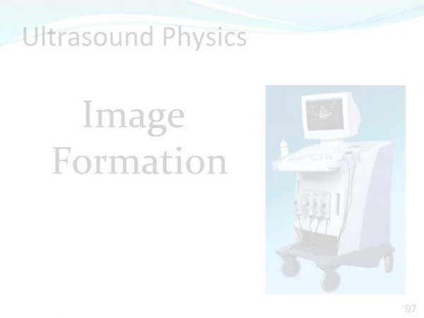
Components Of B mode – Real time machine TIME IS OF THE ESSENCE