Download
1 / 25
270 likes | 310 Views
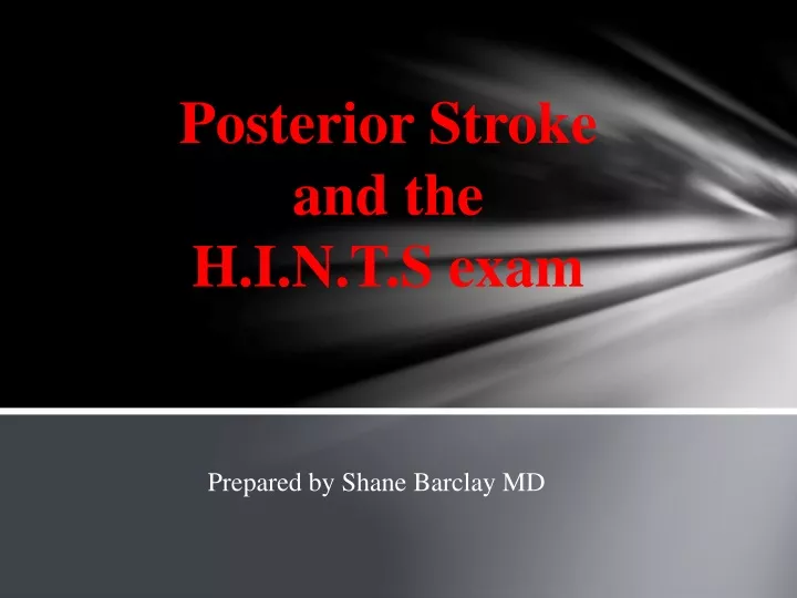
Posterior Stroke and the H.I.N.T.S exam. Prepared by Shane Barclay MD. Posterior Stroke Presentation. Often patients will only present with one symptom: Vertigo The differential is ‘peripheral’ causes of vertigo versus ‘central’ causes. Definition of Vertigo.

E N D
Posterior Strokeand the H.I.N.T.S exam Prepared by Shane Barclay MD
Posterior Stroke Presentation • Often patients will only present with one symptom: • Vertigo • The differential is ‘peripheral’ causes of vertigo versus ‘central’ causes.
Definition of Vertigo • Perception of movement (rotational or otherwise) where no movement exists • Pathophysiology • Mismatch or asymmetric activity of visual, vestibular, and/or proprioceptive systems • Must distinguish peripheral from central cause • Peripheral: 8th CN, vestibular apparatus • Central: Brainstem, cerebellum
Case • 86 year old woman presented with a 2 day history of fairly sudden onset severe vertigo and nausea. No vomiting. One to two weeks prior described a mild viral URTI with some sinus ‘fullness’. • PHx: HTN – controlled • Meds: HCTZ • Exam: CN normal, finger to nose normal, heel/shin normal. Strength and reflexes normal. CV/chest benign. Dix-Halpike exam non conclusive. Slight ataxia on walking. • Labs – CBC, glucose, lytes, GFR – normal.
NeurologyJuly 8, 2014 • “Frequency of False-Negative MRIs and non-lacunar infarcts” • Saber Tehrani AS et al. • 105 patients over 13 yrs were reviewed. All presented with acute vestibular syndrome (days to weeks of continuous vertigo, nausea or vomiting, head-motion intolerance, gait unsteadiness and nystagmus). • Early MRI (within 48 hrs of symptoms) was 47% sensitive for detecting acute infarcts of < 10 mm (most involving the inferior cerebellar peduncle or lateral medulla) • and 92% sensitive for infarcts > 10 mm • Detailed beside exam including HINTS was > 99% sensitive for diagnosing infarcts of all sizes. (HINTS was false negative in only one case)
Differentiating Central versus Peripheral Vertigo • PERIPHERAL • Benign Positional Vertigo • Migranous Vertigo • Vestibular Neuritis • Meniere’s • Viral Labyrinthitis • Drug Toxicity
Differentiating Central versus Peripheral Vertigo • PERIPHERALCENTRAL • Benign Positional Vertigo Cerebellar infarct • Migranous Vertigo Vestibulobasilar TIA • Vestibular Neuritis Chiari Malformation • Meniere’s Multiple Sclerosis • Viral Labyrinthitis Neoplasms • Drug Toxicity
Diagnosis • Sensitivity of Studies • Preference is for MRI due to greater sensitivity
HINTS Exam Stroke September 2009 Journal of the American Heart Association HINTS to Diagnose Stroke in the Acute Vestibular Syndrome: Three-Step Bedside Oculomotor Examination More Sensitive Than Early MRI Diffusion-Weighted Imaging Jorge C. Kattah, Arun V. Talkad, David Z. Wang, Yu-Hsiang Hsieh and David E. Newman-Toker
Diagnosis Test Sensitivity
HINTS Exam • 3 Components 1. Head Impulse test of vestibulo-ocular reflex function 2. Observation for Nystagmus in primary, right, and left gaze 3. Alternate cover Test for Skew deviation.
HINTS Exam • HEAD IMPULSE (or Head Thrust) • Have patient fix their eyes on your nose • Move their head in the horizontal plane to the left and right. • When the head is turned towards the normal side the vestibular ocular reflex remains intact and eyes continue to fixate on the visual target • When the head is turned towards the affected side, the vestibular ocular reflex fails and the eyes make a corrective saccade to re-fixate on the visual target. It is reassuring if the reflex is abnormal(due to dysfunction of the peripheral nerve) ie abnormal means it is a peripheral cause of vertigo.
HINTS Exam • NYSTAGMUS • Peripheral causes of vertigo (ie BPV) can give HORIZONTAL nystagmus but ONLY in one direction. Move the head right, left or up and down and the nystagmus will ONLY be in one direction. • However if you have the patient look to the left and there is left beating nystagmus and then have the patient look to the right and there is right beating nystagmus, that is known as direction changing nystagmus and that is BAD. ie occurs with central cause of nystagmus. • Vertical nystagmus is always BAD.
HINTS Exam • TEST of SKEW • Skew is also known as vertical dysconjugate gaze and is a sign of a central lesion. • 1. Have pt look at your nose with their eyes and then cover one eye • 2. Then rapidly uncover the eye and quickly look to see if the eye moves to re-align. • 3. Repeat on each eye • (4. or if pt complains of binocular diplopia that is a positive test too)
SummaryPatient presents with Continuous Vertigo and no hearing loss. • 1 Head Impulse • Normal patient, eyes will remain fixed on the target (your nose) • Peripheral Vertigo Pt – rapid rotation of the head toward the affected side will result in loss of fixation and movement of the eyes away from the target. • With Central Vertigo, there is typically NO corrective saccade. • i.e. you want there to be saccade motion
Summary Patient presents with Continuous Vertigo and no hearing loss. • 2. Nystagmus • - Normal Pt’s will have NO nystagmus • - Pt’s with peripheral vertigo cause will have • unidirectional, horizontal nystagmus • - Pt’s with central vertigo can have rotatory or vertical • nystagmus, or direction changing nystagmus (right • beating nystagmus when looking right and left beating • nystagmus when looking left) • i.e. you want there to be unidirectional, horizontal • nystagmus.
Summary Patient presents with Continuous Vertigo and no hearing loss. • 3. Test of Skew • - Normal Pt’s will have no skew deviation. • - Pt’s with peripheral vertigo will also not have any • skew deviation • - Pt’s with central vertigo will have misalignment and • therefore as the cover is moved off from the eye, • a slight correction (up or down) will occur. • i.e. you want the patient to NOT have any skew deviation.
Summary Patient presents with Continuous Vertigo and no hearing loss. • So, you can rule out a central cause of vertigo if: • Pt has no corrective saccade with head impulse • Pt has unidirectional horizontal nystagmus. • Pt has no skew deviation. • If the patient has any of the following along with suggestive history, they should be admitted for further evaluation (MRI) for possible central stroke: • Pt has no corrective saccade with head impulse. • Pt has rotatory or vertical nystagmus or direction changing nystagmus • Pt has misalignment and correction of eyes with uncovering of the eye.
HINTS – Summary • Head Impulse • You want the Head Impulse test to be ABNORMAL to reassure you the patient has a peripheral cause of vertigo. • Nystagmus • You want the nystagmus to be fast beating in ONLY ONE DIRECTION to reassure you the patient has a peripheral cause of vertigo. • Test of Skew • You want PERFECT VERTICAL ALIGNMENT of the eyes to reassure you the patient has a peripheral cause of vertigo
Head Thrust TestPt with peripheral cause of vertigo – there is corrective saccade
HINTS Exam • If ANY one of the HINTS exam components is positive, the patient needs a neurological consult/MRI. • A positive HINTS exam: 100% sensitive and 96% specific for the presence of a central lesion.