Download
1 / 42
470 likes | 1.27k Views
Sinusitis. Sinusitis is an extremely common part of the common cold syndrome RVs have been detected in 50% of adult patients with sinusitis by RT-PCR of maxillary sinus brushings or nasal swabs

E N D
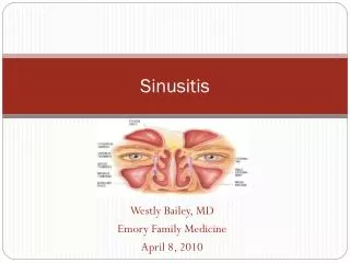
Sinusitis Sinusitis is an extremely common part of the common cold syndrome RVs have been detected in 50% of adult patients with sinusitis by RT-PCR of maxillary sinus brushings or nasal swabs Frequency of association of RV infection with sinusitis suggests that common cold could be considered a rhinosinusitis
Sinusitis Signs and symptoms Patient may complain of a ‘feeling of fullness’ and pressure over the involved sinuses, nasal congestion, and purulent nasal discharge Other associated symptoms include sore throat, malaise, low grade fever, headache, toothache, and cough >1 weeks duration Symptoms may last 10 – 14 days
Sinusitis Diagnosis Based on clinical signs and symptoms Physical examination may reveal patient described symptoms – palpate over sinuses, observe for structural abnormalities such a deviated nasal septum Sinus radiographs may reveal cloudiness and air fluid levels Limited coronal CT are more sensitive to inflammatory changes and bone destruction
Sinusitis Management/Treatment 2/3 of untreated patients will improve symptomatically within 2 weeks Antibiotics may be appropriate in certain patients Supportive therapy such as humidification, antihistamines, analgesics, and/or vasoconstrictors may relieve congestion and fullness OTC decongestant sprays for use of more than 5 days duration should be discouraged
Pharyngitis Fewer than 25% of patients with a sore throat have true pharyngitis Primarily seen in 5 – 18 year old population, it is common in adult women Most common cause is viral; most common agent is rhinovirus; Self-limiting; usually lasts 3-4 days Group A, beta-hemolytic streptococcus is the primary bacterial pathogen in 1/3 cases Early detection reduces incidence of acute rheumatic fever and post streptococcal pharyngitis
Pharyngitis • Sore throat is the prominent symptom • Erythema • Swelling of the affected tissues • Exudates: inflammatory cells overlaying mucous • membranes • Low-grade fever, mild general symptoms • Difficult to differentiate from streptococcal infection Caused by the same viruses that cause common cold and Adenovirus, Enteroviruses and Influenza virus.
Viral Causes of Pharyngitis Rhinoviruses Adenoviruses Coronaviruses Epstein-Barr Virus Herpes Simplex Virus Parainfluenza Viruses Respiratory Syncytial Virus Influenza Viruses Coxsackie Viruses
Adenoviruses 51 serotypes • Immunity correlates with the presence of type- specific • neutralizing antibodies • Endemic or epidemic, often during summer • Incubation period 4-7 days • Moderate to severe pharyngitis, sometimes exudative • Fever and systemic symptoms • Rhinitis and follicular conjunctivitis are common
Adenovirus 51 serotypes Pharyngo-conjunctival fever sporadic or epidemic association with swimming pools • Epidemic acute respiratory disease • in military recruits • pneumonia in 10-20% • Pneumonia in immunocompromised patients • BMT recipients: mortality 60% • Nosocomial transmission: • epidemic keratoconjunctivitis
Pathogenesis Epithelial cells are the primary target. E1B and E4 proteins inhibit transport of host mRNA from the nucleus to the cytoplasm causing cell death The penton protein has been shown to be directly toxic to cells and it has been found in the blood of several fatal cases of adenoviurs pneumonia.
Entry by the mouth, the nasopharynx or via the conjunctiva. The lower stereotypes (1,2,5 and 6) are ubiquitous particularly in young children Endemic spread takes place by the fecal oral route to new pools of susceptible infants and children.
May be transmitted in swimming pools, via medical equipment (tonometer), and via respiratory droplets. Site of initial replication is commonly the oropharynx and spread is mostly local. Virermic spread is rare. Latency has been shown to be common among humans (in tonsils and adenoids)
Adenovirus Clinical Syndromes They infect the respiratory tract as well as the eye, gastrointestinal tract, urinary bladder, and the liver. On occasions, these viruses may cause disease in other organs such as CNS and the pancreas. Most human disease is associated with only one-third of the serotypes. Many adenovirus infections are subclinical
Respiratory Disease Endemic Adenovirus Respiratory Infections of young children - Represent5% of the acute respiratory disease in children(<5y) most commonly as pharyngitis or pharyngoconjunctival fever - Most common serotypes are 1,2,5 and 6 and occasionally 3, 4 and 7. - Responsible for 10% of the pneumonias of childhood. - Most patients recover but epidemics of adenovirus 7 have resulted in considerable mortality.
Acute Respiratory Disease Primarily affects military recruits (types 4, 7 and occasionally 3). Frequently occurs under conditions of fatigue and crowding. Characterized by fever, pharyngitis, cervical adenitis, cough, hoarseness and rhinitis. Some cases have had a fatal outcome (pneumonia).
Pertussis – like syndrome - It is associated with adenovirus type 5. Infections of the Eye - Acute follicular conjunctivitis types 3 and 7 but other types (1,2,4,6,9,10,15,17,20,22) have been incriminated.
Epidemic Keratoconjunctivitis - Types 8, 11, 19 and 37. - Followed by corneal subepithelial infiltration which may persist for a long period but it resolves completely with return of visual acuity to normal. - Outbreaks can be traced to eye clinics where an instrument (Tonometer) or a solution acts as a vehicle.
Viral Causes of Pharyngitis Rhinoviruses Adenoviruses Coronaviruses Epstein-Barr Virus Herpes Simplex Virus Parainfluenza Viruses Respiratory Syncytial Virus Influenza Viruses Coxsackie Viruses
Laryngotracheo Bronchitis (Croup) - An acute viral inflammation of larynx, trachea, and bronchi that is common in young children. - It is often preceded by a "cold". - Accompanied by pyrexia, hoarseness, croaking cough, stridor, restlessness (respiratory insufficiency). - Can be fatal - i.e. life-threatening disease.
Acute Bronchitis Inflammation of bronchi, accompanied by fever, cough, wheezing and "noisy chest". Respiratory virus infection associated with cough Influenza virus: 75%–93% of cases Adenovirus: 45%–90% RVs: 32%–60% Coronaviruses: 10%–50% 40% of nonasthmatic patients with acute bronchitis had FEV180% of predicted Bronchial reactivity remained increased up to 5 weeks after an episode of acute bronchitis
Acute Bronchiolitis Inflammation of terminal bronchioles in young children. - Bronchiole diameter is larger during inspiration than during expiration and this leads to hyperinflation of air sacs distal to bronchiole. - Complete plugging of bronchiole with air resorption leads to collapse. These features can be seen on x-ray. - These changes cause respiratory embarrassment and can be life-threatening. - Clinically, there is fever, rapid respiration, exhausting cough and wheezing.
Pneumonia & Bronchopneumonia - Acute respiratory disease accompanied by fever, restlessness and cyanosis. Often not much clinical "consolidation". Again, can be life-threatening.
Causative Agents Paramyxoviruses - Parainfluenza viruses - Respiratory Syncytial Virus (RSV) - Measles virus Influenza Coronaviruses Adenoviruses Enteroviruses Rhinoviruses
Pathogenesis and Pathology Initially, the mucous membranes of the nose and throat are involved. Obstruction of the paranasal sinuses and eustachian tubes may also occur. Many patients with mild disease may have limited involvement of the bronchi as well. In more extensive infections there is a tendency for HPIV-1 and 2 to involve the larynx and upper tarchea, resulting in croup.
Such infections may extend also to the lower trachea and bronchi, with accumulation of inspissated mucous and resultant atelectasis and pneumonia. When HPIV-3 produces severe disease, infection of the small air passage is likely with the development of bronchopneumonia, bronchiolitis, or bronchitis. Lower respiratory tract involvement also occurs commonly during primary HPIV-1 and 2 infection; about 25% of primary infections produce bronchitis or pneumonia.
The mechanisms responsible for localization and severity of human parainfluenza viruses' disease are not known. Severe respiratory tract disease caused by HPIV1, 2, and 3 generally occurs in the first 3-5 years of life. Primary infections and reinfections occur and most persons have had primary infections before the age of 5 years.
Clinical Features Most infections are asymptomatic, especially in older children and adults. The incubation period is 2-6 days. Fever and a spectrum of respiratory infections are caused by HPIVs; rhinorrhea/rhinitis, pharyngitis, croup, bronchiolitis and pneumonia.
In children, the most common type of illness consists of rhinitis, pharyngitis, and bronchitis, usually with fever. Severe acute laryngotracheobronchitis (Croup) is noted in only 2-3% of primary HP1V1 or 2 infections. When croup develops, the initial symptoms of rhinitis, pharyngitis, fever, and cough progress.
After several days, subglottic region becomes narrower, the cough worsens and becomes brassy, “seallike”, or barking, with hoarseness and stridor. At this stage, most children recover uneventfully after 24-48 hours. In some children, however, air hunger develops, with cyanosis, sternal and intercostal retraction, and progressive airway obstruction.
HP1V3 is an important cause of bronchiolitis in young infants and children below 2 years of age. When bronchiolitis or pneumonia develops, fever persists and the cough progresses and becomes somewhat productive. It is accompanied by wheezing, tachypnea, retraction, and in severe cases cyanosis.
A combined bronchopneumonia-croup syndrome occurs in some patients. Rarely, parainfluenza viruses are associated with otitis media, parotitis, and aseptic meningitis. Prolonged (persistent) and particularly severe infections are known to occur in the immunocompromised
RSV is the most important cause of viral lower respiratory tract disease in infants and children worldwide. RSV is also an important agent of disease in immunosuppressed adults and the elderly. RSV grows poorly in tissue culture and most experimental animals, does not shut off host macromolecular synthesis, and it is unstable. Consequently, research on RSV was impeded.
RSV survives on surfaces for up to 6 hours and on gloves for less than 2 hours. The virus loses activity with freeze-thaw cycles, in acidic conditions and with treatment by disinfectants. Classified into two types, RSV-A and RSV-B, on the basis of variation in the G glycoprotein RSV utilizes ICAM-1 as its receptor.
Pathogenesis RSV is transmitted via large droplets, through fomites and via hands The mechanism of virus spread from upper to lower respiratory tract is assumed to be via the respiratory epithelium or through aspirated secretions. The virus is capable of cell-to-cell spread without emergence into the extracellular fluid. Viremia has not been described during infection of normal infants and children
Pathogenesis RSV causes the release of Interleukins Leukotrienes Chemokines This results in inflammation and tissue damage Presence of eosinophils and eosinophilic cationic protein in blood is associated with recurrent wheezing episodes post RSV infection
Pathogenesis Bronchiolitis Virus induced necrosis of bronchiolar epithelium Hypersecretion of mucous Round cell infiltration and edema of the surrounding submucosa This leads to Formation of mucous plugs Hyperinflation/collapse of distal airways Can also result in interstitial pneumonia Infants are particulary at risk due to small size of normal bronchioles
Pathology Lower respiratory tract involvement (signs) usually appear 1-3 days after the onset of illness (rhinorrhea) Inflammatory infiltration, edema, and excessive mucous production cause obstruction of small bronchioles, with either collapse or emphysema of distal portions of the airway. In those instances in which pneumonia occurs, the interalveolar walls thicken as a result of mononuclear cell infiltration, and the alveolar spaces may fill with fluid.
There is usually a patchy appearance of these pathologic changes, even though disease may be widespread. Severe infections are observed in: - Preterm infants (<35 weeks gestational age) - Those with chronic lung disease - Those with cyanotic congenital heart disease - Immunocompromised hosts.