Download
1 / 15
160 likes | 485 Views
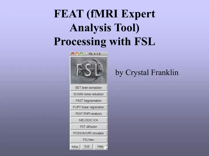
FEAT (fMRI Expert Analysis Tool) Processing with FSL. by Crystal Franklin. Preparing Data for fMRI Analysis. Retrieve data from the archive get_mri- archive contains fmri, T1, and 3d anatomical /archive/tim?_data0/mri_archive Convert images to NIFTI format

E N D
FEAT (fMRI Expert Analysis Tool) Processing with FSL by Crystal Franklin
Preparing Data for fMRI Analysis • Retrieve data from the archive • get_mri- archive contains fmri, T1, and 3d anatomical • /archive/tim?_data0/mri_archive • Convert images to NIFTI format • Run script 4dtonifti on epi data • 4dtonifti session# taskdescription • E.g. 4dtonifti EH0154 fing • Use MANGO to open and save T1 and 3D in NIFTI format • convert2nii • BET • All anatomical images have to be run through BET before using FSL. Form from MRI Operators.
FEAT FMRI Analysis • Balloon Help – Hold mouse arrow on the different options, it will give you helpful information about what the option does. • Progress Watcher – Opens a html page which allows you to see the progress of your analyses. • Brain/Background Threshold- percent of maximum input image intensity
FEAT FMRI Analysis • Number of analyses – Select the number of scans to be analyzed, then select the 4D nifti files • Total Volumes – number of image volumes (time points) • Delete Volumes – The number of scans you would like to delete at the beginning because steady-state has not been reached. • TR – Time from the start of one volume to the start of the next • High Pass Filter Cutoff – the longest temporal period you will allow
FEAT FMRI Analysis • Motion Correction – MCFLIRT which uses FLIRT (FMRIB’s Linear Registration Tool) • Slice timing correction – Corrects for the fact each slice is taken at slightly different times • Not necessary for long epochs. • BET Brain Extraction – A brain mask is created from the first volume in the FMRI data. • Spatial Smoothing FWHM (mm) – This reduces noise without reducing valid activations; by default the smoothing is set at 5mm. • Intensity Normalization – Forces every FMRI volume to have the same mean intensity, by default this is turned off. • Temporal Filtering – Highpass filtering removes baseline drift. Lowpass filtering reduces random noise (can be ran from command line using fslmaths).
FEAT FMRI Analysis • FEAT uses GLM (General Linear Modeling) • http://www.fmrib.ox.ac.uk/fsl/feat5/glm.html • FILM Prewhitening - Makes the statistics valid and maximally efficient • Model Setup: • Model Setup Wizard • Allows you to setup simple experimental designs. • Full Model Setup • You will need to give a text file containing ones and zeros for each explanatory variable.
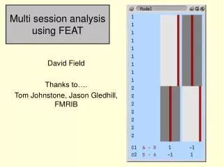
FEAT FMRI Analysis Contrasts • Original EVs- Explanatory Variables • e.g. Right Finger Tapping • Basic Shape- design matrix • Custom (3 Column Format)- Most commonly used. 3-Column Text File **Be sure to remember the order you enter your EVs, you will need it for the contrasts**
FEAT FMRI Analysis Design Matrix/Efficiency Eigenvalues % change required for each contrast to pass specified z-threshold
FEAT FMRI Analysis Post-stats • The data is usually run with the default threshold of Z > 2.3.
FEAT FMRI Analysis Registration Initial Structure – T1 weighted image, if acquired. Main Structure – 3D anatomical image. Standard Space – Should be an image in Talairach space; we usually use the Colin Brain. **You can save the design you setup so that you can just load the design.fsf file without having to setup the contrast and EVs again**
FEAT FMRI Analysis Results • For each analyses ran through FEAT an output directory is created with the extension “.feat” • This folder you will contain all statistical images and files. • e.g. EH0154_fing.feat • The results can be viewed by clicking on the report.html. • This will included the Pre-stats, Stats, Post-Stats, Registration, and Log.
FEAT FMRI Analysis Results
FEAT FMRI Analysis Results
FEAT FMRI Analysis Results
FEAT FMRI Analysis Results