Download
1 / 53
530 likes | 740 Views
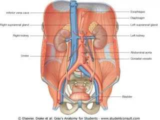
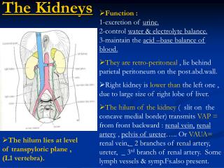
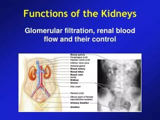
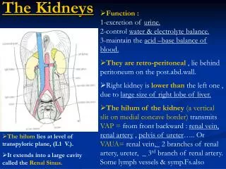
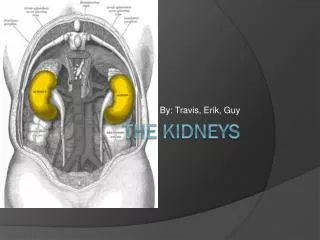
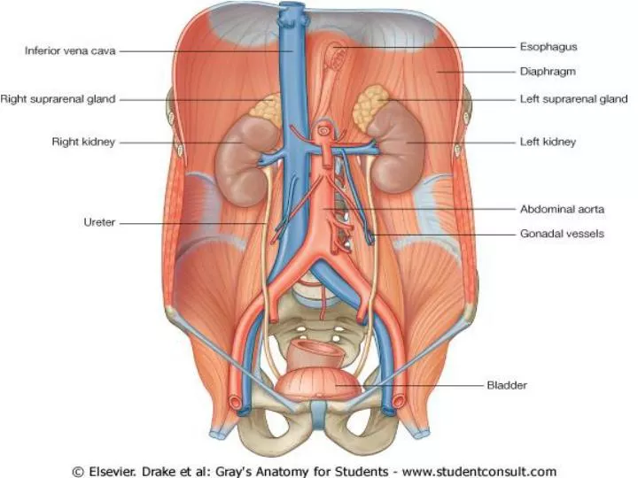
General Organization Of The Kidneys. Location : Posterior wall of the abdomen, outside the peritoneal cavity Size :150 grams, size of clenched fist Medial side: Indented region called the hilum The kidney is surrounded by a tough, fibrous capsule.

E N D
General Organization Of The Kidneys Location : Posterior wall of the abdomen, outside the peritoneal cavity Size :150 grams, size of clenched fist Medial side: Indented region called the hilum The kidney is surrounded by a tough, fibrous capsule
Structures passing through hilum are • Renal arteries • Renal vein • Ureter • Lymphaticsand nerves
Renal Blood Supply • 22 percent of the cardiac output • 1100 ml/min
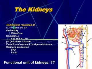
The Nephron(Functional Unit) • Each kidney contains 1million nephrons • Each nephron contains 1) tuft of glomerular capillaries called the glomerulus 2) long tubule in which the filtered fluid is converted into urine • The glomerulus contains a network of branching and anastomosing glomerular capillaries
Glomerulus is encased in Bowman’s capsule • There are two capillary beds • Glomerulus (hydrostatic pressure is 60 mm Hg • Peritubular capillary bed (hydrostatic pressure is13mm Hg)
Fluid filtered from the glomerular capillaries flows into Bowman's capsule and then into the proximal tubule which lies in the cortex of the kidney • From the proximal tubule fluid flows into the loop of Henlewhich dips into the renal medulla. • Each loop consists of a descending and an ascending limb
The walls of the descending limb and the lower end of the ascending limb are very thin and are called the thin segment of the loop of Henle • After the ascending limb of the loop returns back to the cortex its wall becomes thicker and it is referred to as the thick segment of the loop of Henle
At the end of the thick ascending limb is a short segment that has in its wall a plaque of specialized epithelial cells known as the macula densa • Beyond the macula densafluid enters the distal tubule which like proximal tubule lies in the renal cortex • This is followed by the connecting tubule and the cortical collecting tubule which lead to the cortical collecting duct
The initial parts of 8 to 10 cortical collecting ducts join to form a single larger collecting duct that runs downward into the medulla and becomes the medullary collecting duct • The collecting ducts merge to form progressively larger ducts that eventually empty into the renal pelvis through the tips of the renal papillae • In each kidney there are about 250 of the very large collecting ducts each of which collects urine from about 4000 nephrons
Cortical Nephrons • Nephrons whose glomeruli are located in the outer cortex are called cortical nephrons • They have short loops of Henlethat penetrate only a short distance into the medulla • In cortical nephrons the tubular system is surrounded by an extensive network of peritubularcapillaries
Juxtamedullary Nephrons • 20 to 30% nephrons have glomeruli that lie deep in the renal cortex near the medulla and are called juxtamedullarynephrons • They have long loops of Henle that dip deeply into the medulla • In juxtamedullarynephrons long efferentarteriolesextendfrom the glomeruli down into the outer medulla and then divide into specialized peritubular capillaries called vasa recta that extend downward into the medulla lying side by side with the loop of Henle
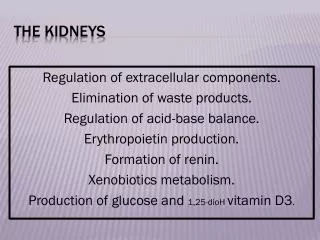
FUNCTIONS OF KIDNEYS • Maintaining water balance in the body • Maintaining the proper osmolality of the body fluids • Regulating the concentration of most of the ECF ions • Excretion of metabolic waste products (urea, uric acid, creatinine, bilirubin, hormone metabolites) • Excretion of foreign compounds (drugs, food additives)
FUNCTIONS (cont) • Production of Renin (enzymatic hormone) • Production of Erythropoietin • Gluconeogenesis (during prolonged period of fasting) • Activation of vitamin D (alpha-1-hydroxylase) • Regulation of acid-base balance (by adjusting urinary out put of hydrogen ions and bicarbonate ions, excretion of sulfuric acid and phosphoric acid) • Regulation of arterial pressure
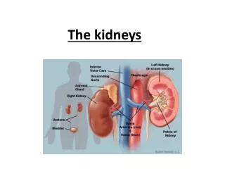
On bisection from top to bottom two regions are seen • Outer cortex and inner medulla Medulla is divided into • 8 to 10 Renal pyramids (cone shaped). The base of each pyramid originates at the border between cortex and medulla and terminates into Papilla • Papilla projects into renal pelvis(funnel-shaped continuation of upper end of ureter) • Renal pelvis is divided into 1. major calyx 2. minor calyx
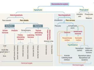
Juxtaglomerular Apparatus (complex) • The juxtaglomerular complex consists of macula densa cells in the initial portion of the distal tubule and juxtaglomerular cells in the walls of the afferent and efferent arterioles • The macula densa is a specialized group of epithelial cells in the distal tubules that comes in close contact with the afferent and efferent arterioles • The macula densa cells sense sodium levels and volume load of the filtrate and cause the release of Renin from JG cells
Micturition • Micturition is the process by which the urinary bladder empties when it becomes filled • First the bladder fills progressively until the tension in its walls rises above a threshold level • In the second step which is a nervous reflex called the micturition reflex that empties the bladder or if this fails at least causes conscious desire to urinate
The micturition reflex is an autonomic spinal cord reflex • It can also be inhibited or facilitated by centers in the cerebral cortex or brain stem
Physiologic anatomy of bladder • Two major parts 1) Body in which urine collects 2) Neck funnel shaped part which connects with the urethra
The smooth muscle fibers of the bladder is called detrusor muscles • The bladder neck (posterior urethra) is 2 to 3 centimeters long. The muscle in this area is called the internal sphincter • Beyond the posterior urethra the urethra passes through the urogenital diaphragm which contains a layer of muscle called the external sphincter of the bladder. This muscle is voluntary skeletal muscle
Innervation Of Bladder Spinal cord segments S-2 and S-3 -----sacral plexus-----pelvic nerves----both sensory nerve fibers and motor nerve fibers Sensory fibers detect the degree of stretch Motor nerve fibers are parasympathetic fibers
Skeletal motor fibers through the pudendal nerves----external sphincter
The bladder receives sympathetic innervation from the sympathetic chain through the hypogastricnerves connecting mainly with the L2 segment of the spinal cord These sympathetic fibers stimulate mainly the blood vessels and have little to do with bladder contraction
Urine flowing from the collecting ducts into the renal calyces stretches the calyces and increases their inherent pacemaker activity which in turn initiates peristaltic contractions that spread to the renal pelvis and then downward along the length of the ureter forcing urine from the renal pelvis toward the bladder
Peristaltic contractions in the ureter are enhanced by parasympathetic stimulation and inhibited by sympathetic stimulation • Vesicoureteral reflux • Ureterorenal reflex The ureters are well supplied with pain nerve fibers
The smooth muscle of the bladder shows the property of tone, accommodation (adaptation, receptive relaxation) and rhythmic contraction
When there is no urine in the bladder, the intravesicular pressure is about 0 • When 30 to 50 milliliters of urine have collected the pressure rises to 5 to 10 centimeters of water • Additional urine-200 to 300 milliliters can collect with only a small additional rise in pressure • Beyond 300 to 400 milliliters collection of more urine in the bladder causes the pressure to rise rapidly
Micturition contractions are the result of stretch reflex initiated by sensory stretch receptors in the bladder wall • Sensory signals from the bladder stretch receptors are conducted to the sacral segments of the cord through the pelvic nerves and then back again to the bladder through the parasympathetic nerve fibers of the pelvic nerves
When the bladder is only partially filled the micturition contractions relax spontaneously after a fraction of a minute and pressure falls • As the bladder continues to fill the micturition reflexes become more frequent and cause greater contractions of the detrusor muscle
Once micturition reflex begins it is self-regenerative • Initial contraction of the bladder activates the stretch receptors which increases the sensory impulses going from the bladder to the spinal cord • This increases reflex contraction of the bladder • The cycle is repeated again and again until the bladder has reached a strong degree of contraction • After few seconds to more than a minute the self-regenerative reflex begins to fatigue causing the bladder to relax
Once micturition reflex has occurred but has not succeeded in emptying the bladder the nervous elements of the reflex remain in inhibited state for a few minutes to 1 hour or more before another micturition reflex occurs
Role of the Higher Centers The micturition reflex is an autonomic spinal cord reflex but it can be inhibited or facilitated by centers in the brain. The centers include • Strong facilitative and inhibitory centers in the brain stem located mainly in the pons • Centers located in the cerebral cortex that are mainly inhibitory but can become excitatory
If the micturition reflex becomes powerful enough it causes another reflex which passes through the pudendal nerves to the external sphincter to inhibit it • If this inhibition is more potent in the brain than the voluntary constrictor signals to the external sphincter urination will occur • If not urination will not occur until the bladder fills still further and the micturition reflex becomes more powerful
The higher centers keep the micturition reflex partially inhibited except when micturition is desired • The higher centers can prevent micturition, even if the micturition reflex occurs by tonic contraction of the external bladder sphincter until a convenient time presents itself • When it is time to urinate the cortical centers can facilitate the sacral micturition centers to help initiate a micturition reflex and at the same time inhibit the external urinary sphincter so that urination can occur
Voluntary Urination • First a person voluntarily contracts the abdominal muscles which increases the pressure in the bladder and allows extra urine to enter the bladder neck and posterior urethra under pressure thus stretching their walls • This stimulates the stretch receptors which excites the micturition reflex