Download
1 / 52
520 likes | 791 Views
Eyelid Cancer and Reconstruction. Laurence Z. Rosenberg,M.D. Southeastern Plastic Surgery. Benign Lesions. Chalazion. Benign Lesions. Chalazion Caused by a blocked duct from a meibomian gland This is not a sty (glands of Zeis) Initial treatment warm compresses

E N D
Eyelid Cancer and Reconstruction Laurence Z. Rosenberg,M.D. Southeastern Plastic Surgery
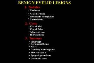
Benign Lesions Chalazion
Benign Lesions Chalazion Caused by a blocked duct from a meibomian gland This is not a sty (glands of Zeis) Initial treatment warm compresses May require surgical excision
Benign Lesions Chalazion Cautious observation for a limited time. If the lesion is not getting better, refer or do a biopsy. If you excise the lesion, always send the specimen to pathology
Benign Lesions Trichoepithelioma
Benign Lesions Trichoepithelioma Benign lesion, often develop after puberty May be numerous Obsevation is indicated
Benign Lesions Trichoepithelioma Desmoplastic trichoepithelioma may resemble a basal cell carcinoma If there is change, never hesitate to biopsy
Benign Lesions Verruca
Benign Lesions Verruca Caused by Human Papilloma Virus. Over 150 strains Filliform warts: long thin lesions on the face Different from genital warts
Benign Lesions Verruca Usually clear up in children without treatment May be more persistent in adults Multiple treatments, but on eyelid, careful excision o cauterization 20% recurrence
Benign Lesions Inclusion Cyst
Benign Lesions Inclusion Cyst Often called a sebaceous cyst, this is a misnomer May resemble a basal cell carcinoma May become inflamed or infected
Benign Lesions Inclusion Cyst Usually no treatment is required Often removed for appearance or because of infection Remove entire cyst and punctum if possible.
Benign Lesions Nevus
Benign Lesions Nevus Atypical Nevus Size >5 mm diameter Ill-defined or blurred borders Irregular margin resulting in an unusual shape Varying shades of color (mostly pink, tan, brown, black) Flat and bumpy components
Benign Lesions Nevus Dysplastic Nevi –pathologic diagnosis The lesion may be a junctional naevus or more frequently a compound naevus (the cells are found at the epidermodermal junction and within the dermis). The nevus cells form a row along the dermoepidermal junction (called lentiginous proliferation), with or without nevus cells in nests (called theques).
Benign Lesions Nevus Dysplastic Nevi –pathologic diagnosis These theques are often irregular in size and shape and may 'bridge' or join together. The cells may be odd-looking i.e. they have cytologic atypia, and they may be spindle-shaped (elongated) or epithelioid (resembling epidermal keratinocytes i.e., broad). There may be fibrosis or scarring in the dermis.
Benign Lesions Nevus Dysplastic Nevi –pathologic diagnosis Inflammatory cells may infiltrate the lesion. Associated blood vessels may be increased in number or enlarged.
Benign Lesions Nevus Treatment May be for cosmetic purposes Excision dependent on the degree of atypia (moderate or severe) not an exact science
Malignant Lesions Basal Cell Carcinoma
Malignant Lesions Basal Cell Carcinoma Most common human cancer More common in fair skinned people May be heritable: Basal Cell Nevus Syndrome
Malignant Lesions Basal Cell Carcinoma Nodular BCC Most common type on the face Small, shiny, skin colored or pinkish lump Blood vessels cross its surface May have a central ulcer so its edges appear rolled Often bleeds spontaneously then seem to heal over
Malignant Lesions Basal Cell Carcinoma Superficial BCC Often multiple Anywhere Pink or red scaly irregular plaques Slowly grow over months or years Bleed or ulcerate easily
Malignant Lesions Basal Cell Carcinoma Morpheaform BCC Also known as sclerosing BCC Usually found in mid-facial sites Prone to recur after treatment May infiltrate cutaneous nerves (perineural spread)
Malignant Lesions Basal Cell Carcinoma Pigmented BCC Brown, blue or greyish lesion Nodular or superficial histology May resemble melanoma
Malignant Lesions Basal Cell Carcinoma Basisquamous BCC Mixed BCC and Squamous Cell Carcinoma More Aggressive
Malignant Lesions Basal Cell Carcinoma Treatment Currettage and cautery: Margins unknown Excision: Margins known, but not circumferential Mohs: Best for high risk lesions, most definitive margin assessment Photodynamic Therapy: superficial BCC. Lower Cure Rate Imiquimod: Immune modulator Radiation: May be used in elderly or as adjuvant therapy
Malignant Lesions Squamous Cell Carcinoma
Malignant Lesions Squamous Cell Carcinoma Directly related to UV exposure Smoking Chronic wounds Human Papiloma Virus
Malignant Lesions Squamous Cell Carcinoma Treatment: Surgery Excision Mohs Patient may require assessment of the lymph nodes Large tumors may require pre-operative radiographic imaging
Malignant Lesions Squamous Cell Carcinoma 5% metastasize to other sites more likely in transplant patients, old age, alcoholics etc. May require adjuvant radiation therapy
Malignant Lesions Melanoma
Malignant Lesions Melanoma Cancer of the melanocytes Prognosis dependent of tumor thickness Stage IA: Melanoma <1.0mm Stage IB: Melanoma is <1.0mm with ulceration or Mitoses >1 or > 1.0mm and ≤ 2.0mm Stage IIC: Melanoma > 4.0mm, with Ulceration Stage IIIC: Nodal Involvement or Intransit spread Stage IV: Spread to distant organs
Malignant Lesions Melanoma Stage IA: The 5-year survival rate is around 97%. The 10-year survival is around 95%. Stage IB: The 5-year survival rate is around 92%. The 10-year survival is around 86%. Stage IIC: The 5-year survival rate is around 53%. The 10-year survival is around 40%. Stage IIIC: The 5-year survival rate is around 40%. The 10-year survival is around 24%.
Malignant Lesions Melanoma Stage IV:The 5-year survival rate for stage IV melanoma is about 15% to 20%. The 10-year survival is about 10% to 15%.
Malignant Lesions Melanoma Treatment: Dependent on tumor thickness in-situ 0.5cm < 1.0mm 1cm 1.0 – 2.0mm 1 – 2cm >2.0mm 2cm If the tumor is > 1.0mm thick, or ulcerated or mitotic index ≥ 1 Perform sentinel lymph node biopsy
Reconstruction Mohs defect 45 by 55mm 50% lower Lid 30% Upper lid Resection of lateral canthus Loss of temporal skin
Reconstruction Repair of the eyelid, like all reconstruction: Knowledge of the anatomy Function of the part to be reconstructed Application of technique