Download
1 / 20
200 likes | 411 Views
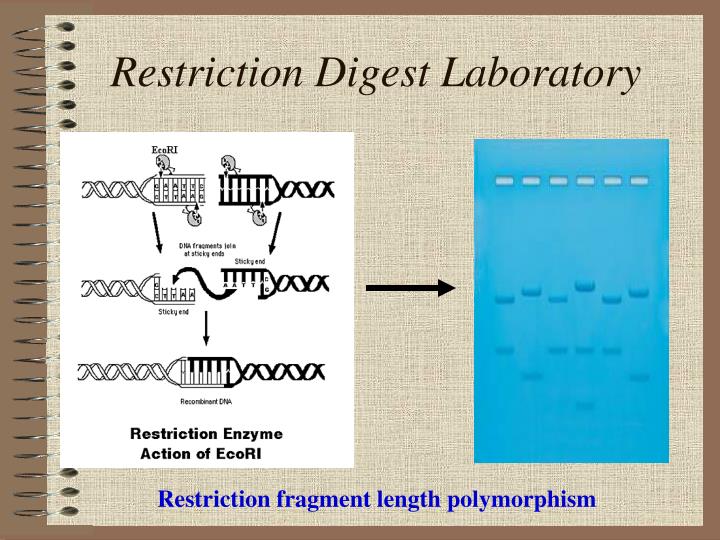
Restriction Digest Laboratory. Restriction fragment length polymorphism. Reminder. You have transformed bacteria with plasmid DNA You have isolated plasmid DNA Today you will perform an RFLP analysis & Confirm your Plasmid Isolation.

E N D
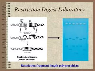
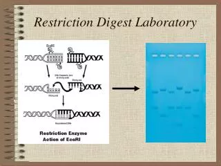
Restriction Digest Laboratory Restriction fragment length polymorphism
Reminder • You havetransformed bacteria with plasmid DNA • You haveisolated plasmid DNA • Today you will perform an RFLP analysis • & Confirm your Plasmid Isolation
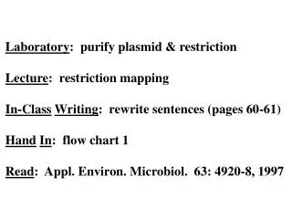
This is the third and final section of your lab report. • Digest plasmid DNA • Determine number of cutting sites • Determine location of cutting sites • Determine size of fragments • Present the “map” of the plasmid in your report The steps in BLUE you will complete outside of class as part of your data analysis.
What is: • A restriction enzyme(s)? • An endonuclease • We will focus on type II. • A restriction digest?
Examples of Restriction Enzymes Links to restriction enzymes: http://www.neb.com/nebecomm/tech_reference/restriction_enzymes/buffer_activity_restriction_enzymes.asp http://www.accessexcellence.org/AE/AEC/CC/re_chart.php http://www.neb.com/nebecomm/EnzymeFinder.asp?
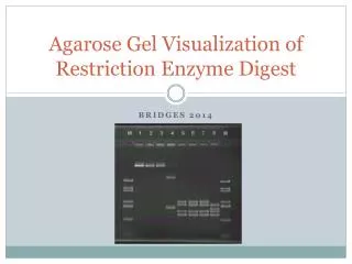
Analysis of Data Allows you to identify sizes of plasmid By comparing migration of digested plasmid To KNOWN SIZES of DNA.
A map gives the size of fragments • A map gives the number and position of cutting sites 60 800 600 JUST AN EXAMPLE Not your map! 1500 Plasmid map 1400
Remember Plasmid is Circular • Circular DNA: the number of fragments=number (N) of cutting sites • versus • Linear DNA: number of fragments=N+1
Linear DNA Plasmid DNA 2 cutting sites 2 fragments 2 cutting sites 3 fragments
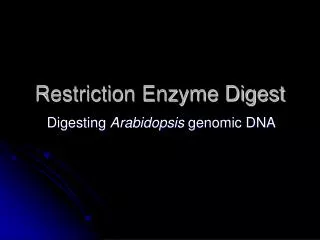
Today’s experiment Restriction of Digest of plasmid DNA using two restriction enzymes.
Please refer to page 10 of the handout(6 groups) • Each Group set up a rack with: • Reaction buffer • water • Plasmid DNA • NotI for AM lab • SfiI for AM lab • Or • AluI for PM lab • HphI for PM lab • Loading Dye • Standard (marker or ladder) DNA • Label four microfuge tubes 1→4 Must keep on ice
Pipette the samples as shown on page in handout—not lab manual.
After you are finished pipetting your samples • Place samples at 37C for 1 hour • After 1 hour you will be ready to load your gel
Restriction Digest • AFTER 1 hour DIGESTION: You must add 5 ul 10X loading dye to your samples (not to the ladder (L)). • Pre-heat all samples including ladder for 3-5 min. at 65C
Gel Electrophoresis • Load 25 ul per well • Run gel at 75 volts until the dye front is approximately half-way down gel. • Take photograph