Download
1 / 35
350 likes | 456 Views
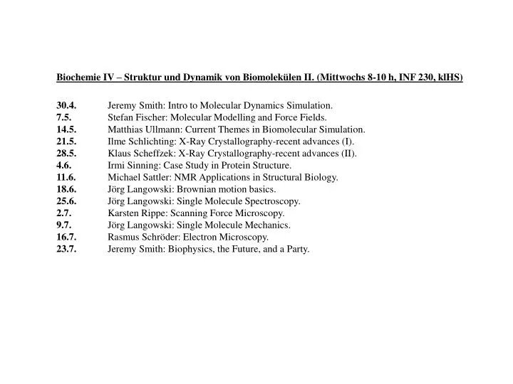
Biochemie IV – Struktur und Dynamik von Biomolekülen II. (Mittwochs 8-10 h, INF 230, klHS) 30.4. Jeremy Smith: Intro to Molecular Dynamics Simulation. 7.5. Stefan Fischer: Molecular Modelling and Force Fields. 14.5. Matthias Ullmann: Current Themes in Biomolecular Simulation.

E N D
Biochemie IV – Struktur und Dynamik von Biomolekülen II. (Mittwochs 8-10 h, INF 230, klHS) 30.4. Jeremy Smith: Intro to Molecular Dynamics Simulation. 7.5. Stefan Fischer: Molecular Modelling and Force Fields. 14.5. Matthias Ullmann: Current Themes in Biomolecular Simulation. 21.5. Ilme Schlichting: X-Ray Crystallography-recent advances (I). 28.5. Klaus Scheffzek: X-Ray Crystallography-recent advances (II). 4.6. Irmi Sinning: Case Study in Protein Structure. 11.6. Michael Sattler: NMR Applications in Structural Biology. 18.6. Jörg Langowski: Brownian motion basics. 25.6. Jörg Langowski: Single Molecule Spectroscopy. 2.7. Karsten Rippe: Scanning Force Microscopy. 9.7.Jörg Langowski: Single Molecule Mechanics. 16.7. Rasmus Schröder: Electron Microscopy. 23.7. Jeremy Smith: Biophysics, the Future, and a Party.
GRAMICIDIN S - cyclo(Leu-DPhe-Pro-Val-Orn)2 - Powerful but nonspecific antimicrobial agent. - Principal target : bacterial or erythrocyte membranes.
Structure- Antimicrobial Activity Relationships: Two basic residues (e.g. Orn) on same face - required. Hydrophobic residues in Leu/Val positions - required. sheet and turns - required. : Sidedness Hypothesis (Schwyzer, 1958, Kato & Izumiya, 1977)
Molecular Dynamics of Gramicidin S in DMSO Backbone: Stays in one conformation Average deviation from NMR: 18o NMR: Xu et al 1995.
Order parameters of the sn-2 chains of DMPC. Hydrated DMPC -Douliez et al 1975 Bound Lipids - Disordered Free lipids - more ordered
STÉPHANIE HÉRY DANIEL GENEST Scattering of X-Rays by Protein Crystals Real Crystal Ideal Crystal + Perturbations =
Molecular Dynamics of Lysozyme Unit Cell Full Trajectory Experimental Rigid-Body Decomposition Rigid-Body Fit (R-factor re: Full Trajectory = 5.3%)
FRANCI MERZEL Protein Hydration. Svergun et al PNAS 1998: First 3Å hydration layer around lysozyme ~10% denser than bulk water
Geometric Rg from MD simulation = 14.10.1Å
(d) (d) Bulk Water d Bulk Water Average Density Present Even if Water UNPERTURBED from Bulk o(d) Bulk Water Radial Water Density Profiles Water Protein o(d) 10% increase o(d)- (d) = Perturbation from Bulk 5% increase
What determines water density variations at a protein surface?
Simple View of Protein Surface (1) Topography h=Surface Topographical Perturbation Protuberance L=3 surface Depression L=17 surface + (2) Electric Field qi qj qk
Surface Topography, Electric Field and Density Variations Low High O High H H High
Conclusions (1) Simulation and Experimental I(q) in Good Agreement (2) First Hydration Layer (0-3Å) ~15% Density Increase of which: - ~10% Unperturbed - ~5% Perturbed Fewer Disorienting Bulk Water Dipoles Water Dipoles Align with Protein E Field Water Density Variations Correlated with Surface Topography and Local E Field from Protein
More Proteins Protein 1 Protein 2 Complex Formation Conformational Change Function
Structures of Macromolecular Complexes • Very few experimentally determined • e.g. antibodies:antigens • ~1000 antibody sequences known • ~100 antibody structures known • ~10 antibody:antigen complex structures known • Can we use calculation?
Homology Modelling Can derive structures for sequences with >20-30% sequence identity when aligned with sequence of known structure.
Structures of Isolated Components? • - crystallography • - NMR • - Homology Modelling • Structure of Complex? • Rigid-Body Shape Complementarity • (based on hydrophobic effect and van der Waals packing) • Conformational Change on Complexation? • Electrostatic Complementarity? • Solvation Effects? • Experiment?
Functional Binding Site on Toxin Red: Affinity Lowered >100-fold Yellow: Affinity Lowered 10-100 fold
Modelling of Isolated Antibody Homology Model of Framework Residues. Complementarity Determining Region Loops (CDRs): (i) Uniform Conformational Searching (ii) Canonical Loop Modelling (iii) Data-Base Searching of Loop Conformations (iv) Molecular Dynamics in vacuo and with solvated CDRs. > 90 models. Clustering and Screening for Consistency with Experimental Antibody Structures. 4 Dynamically Interconvertible Models.
Modelling of Ab:Ag Complex Initial Generation Low -Resolution Shape Complementarity. > 41,585 models Clustering and Screening for: (i) Buried Surface Area. (ii) Electrostatic Complementarity. (iii) Consistency with existing Ab:Ag complex structures. > 18 models. Refinement of Atomic-Detail Models with Molecular Dynamics in Explicit Solvent. 6 Models.
Toxin and M 23 Functional Binding Sites Red - >100 fold affinity loss on mutation Yellow - 10-100 fold affinity loss on mutation
Three Models of Calculated M23 Paratope Red: Residues contacting antigen energy core Yellow: Residues contacting functional epitope
Orientation of toxin on M23 combining site in the two remaining models.
Annexin V - Pathway for Conformational Transition
Charge Transfer in Biological Systems • Ions, Electrons...
NICOLETA BONDAR MARCUS ELSTNER STEFAN FISCHER SANDOR SUHAI Proton Transfer Step #1 in Bacteriorhodopsin