Download
1 / 37
380 likes | 570 Views
Making Zebrafish Glow!. Varun Bhadha Ruth McLaughlin Summer Research Connection The Center of Excellence in Genomic Science 24 July 2009. Photo provided by Frederique Ruf. How can we study early development?. Why study early development?. Developmental diseases

E N D
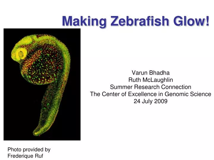
Making Zebrafish Glow! Varun Bhadha Ruth McLaughlin Summer Research Connection The Center of Excellence in Genomic Science 24 July 2009 Photo provided by Frederique Ruf
How can we study early development? Why study early development? • Developmental diseases • Understanding growth and organogenesis • Interesting and exciting • Localized expression • Co-injecting marker • Sub-cellular anatomy
A Solution: Fluorescent Proteins Green Fluorescent Protein jellyfish
Research Goals Generate localized fluorescent markers that will be injected into zebrafish embryos to study embryonic development • Membrane localized protein, cherry-red • Nucleus localized protein, cerulean-blue Zebrafish cell nucleus
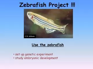
Zebrafish as a Model Organism • Housing and husbandry • * adult fish (3-4cm) can be housed at high density - reduces costs (c/f mice) • * new strains and crosses can be easily bred (paired matings and crosses) • Developmental biology • zebrafish are vertebrates and have a gene complement very similar to humans • gene sequence is mostly done • development is rapid - organogenesis is virtually complete within 3 days • each female can lay >200 eggs per week • Imaging • high-resolution imaging of RNA and protein expression in whole embryos is easy • eggs are transparent, so early developmental processes can be easily visualized • transgenic and mutant lines expressing fluorophores can be easily imaged
chorion cells yolk • 4 cell stage, ~1 hours post fertilization (hpf) • chorion, embryo and yolk are all transparent • cells of the embryo can easily be injected with dyes, gene-specific morpholinos (to inactivate genes), or visualised by microscopy.
3 dpf • organogenesis is almost complete. • the mouth, eye, otic vesicle (ear primordia), gut, liver, somites (muscles) & tail can all be seen. • majority of the embryo is still optically transparent at this age.
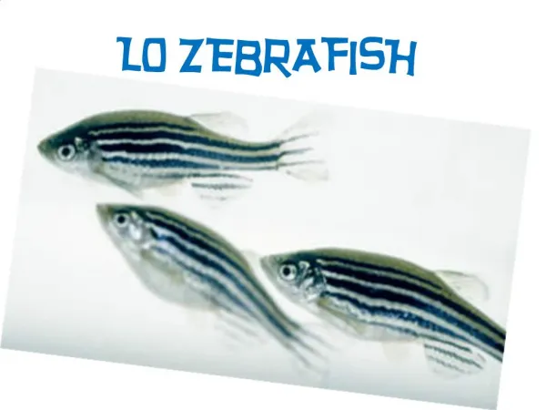
Adult zebrafish (female) 3-4 mpf • No longer transparent. • Sexually mature
Preparation of Genetic Material plasmids Restriction digest Gel extractions Gel electrophoresis ligation Bacterial transformation Isolate new plasmid and clone transcribe inject
DNA Plasmids • Vectors, or plasmids, are circular strands of DNA used by bacteria. • Insert small fragments of DNA into bacterial plasmids that code for the coloring of the nucleus (cerulean-blue) and the cell membrane (cherry-red). • “Cut” or digest our plasmids using restriction enzymes.
Preparation of Genetic Material plasmids Restriction digest Gel extractions Gel electrophoresis ligation Bacterial transformation Isolate new plasmid and clone transcribe inject
Restriction Digest Restriction enzyme cutting sites
Preparation of Genetic Material plasmids Restriction digest Gel extractions Gel electrophoresis ligation Bacterial transformation transcribe Isolate new plasmid and clone inject
Gel Electrophoresis • Separates DNA fragments • Uses an agarose gel and an electrical charge - + Gel box
Gel Electrophoresis (cont.) • Similar to children in a forest with the incentive of a candy bar • Forest = gel • Candy bar = charge • Children = DNA
Preparation of Genetic Material plasmids Restriction digest Gel extractions Gel electrophoresis ligation Bacterial transformation Isolate new plasmid transcribe inject
Gel Extraction • The DNA we need is now inside the gel. To get it out: • Cut DNA from the gel with razor blades • Extract the DNA from the gel piece
Preparation of Genetic Material plasmids Restriction digest Gel extractions Gel electrophoresis ligation Bacterial transformation Isolate new plasmid transcribe inject
Ligation and Transformation • Take the extracted DNA and “paste” together to form the desired plasmid • Insert the DNA into competent bacterial cells (transformation) • Plate the bacteria
Preparation of Genetic Material plasmids Restriction digest Gel extractions Gel electrophoresis ligation Bacterial transformation transcribe Isolate new plasmid and clone inject
DNA Replication and Extraction • Isolate and grow bacterial colonies • Extract the DNA from the grown bacteria • Analyze the data to choose which bacterial DNA to use for the rest of the process
DNA Extraction Data Tube # - DNA Concentration (ng/uL) 1- 56.2 2- 195.6 3- 20.0 4- 189.7 5- 82.8 6- 125.4 7- 428.2 8- 139.1 9- 125.3 10- 154.0 11- 108.4 12- 119.6 13- 159.9 14- 141.9 15- 86.8 16- 59.8
DNA Linearization • The plasmid needs to be turned into a linear strand for transcription
DNA Linearization (cont) Not1 DNA Plasmid Linearized DNA strand Not1 Not1
Preparation of Genetic Material plasmids Restriction digest Gel extractions Gel electrophoresis ligation Bacterial transformation transcribe Isolate new plasmid and clone inject
DNA Strand to RNA • To inject into the embryo, we will need to use RNA • Therefore, we must transcribe RNA from the DNA. https://eapbiofield.wikispaces.com/file/view/c7.17.7b.transcription.jpg
Preparation of Genetic Material plasmids Restriction digest Gel extractions Gel electrophoresis ligation Bacterial transformation transcribe Isolate new plasmid and clone inject
Injecting ~30 min pf 1 cell stage of embryo Single cell • RNA is introduced into the cell by injection at the 1 cell stage. Injection needle Size - 0.7 mm http://images.google.com/imgres?imgurl=http://www.biocompare.com/images/bc/006/ArticleImages/AAA_1.jpg&imgrefurl=http://www.biocompare.com/Articles/TechnicalArticle/1657/Microinjection-Of-DNA,-RNA-And-Tracer-Dyes-Into-Early-Fish-Embryos-from-Eppendorf-AG.html&usg=__mjTx6MdnzrmnbTeV0SbGSsp-E6U=&h=495&w=904&sz=301&hl=en&start=34&tbnid=JZiQGvLg5c2jrM:&tbnh=80&tbnw=147&prev=/images%3Fq%3Dzebrafish%2Binjections%26gbv%3D2%26ndsp%3D21%26hl%3Den%26sa%3DN%26start%3D21%26newwindow%3D1
Conclusions • Genetic material (DNA/RNA) can be manipulated ( as in removed, changed, and inserted) between species. • We have successfully manipulated zebrafish embryos to cause them to fluoresce
Acknowledgements Thank you to… Fraser Laboratory Dr. Aidyl Gonzalez-Serricchio Ilana Solomon Dr. Scott Fraser Kristina L. Hilands Leigh Ann Fletcher SRC Administrators James Maloney Dr. Sherry Tsai Funding Siemens Foundation Howard Hughes Medical Institute Pasadena Independent Schools Foundation Caltech Oak Crest Institute of Science Our Families Our fellow SRC researchers