Download
1 / 24
290 likes | 519 Views
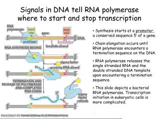
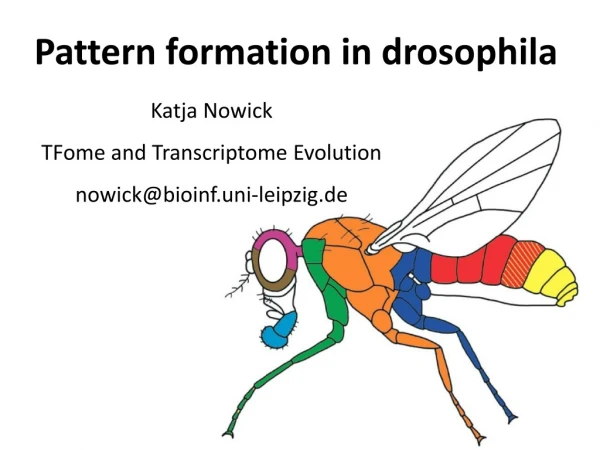
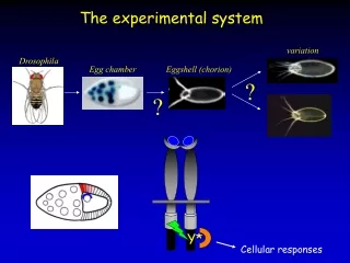
Transcription by Polymerase II in Drosophila. The 3 main phases of the transcription cycle are known as initiation , elongation and termination . Each stage is subject to regulation .

E N D
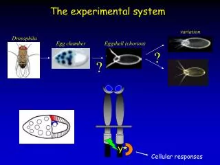
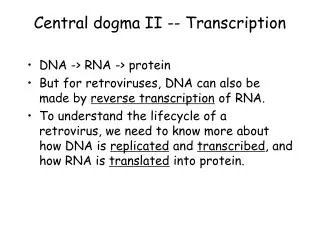
Transcription by Polymerase II in Drosophila The 3 main phases of the transcription cycle are known as initiation, elongation and termination. Each stage is subject to regulation. During transcription initiation, a transcription-competent RNA polymerase complex forms at the promoter and the DNA template is aligned in the active site of the polymerase. The active site is where nucleotides are paired with the template and are joined processively during elongation to produce the RNA transcript. Termination of transcription involves release of the RNA transcript and the dissociation of the transcription complex from the DNA template. Regulation of transcription occurs at the level of RNA polymerase recruitment to the promoter and at the level of elongation. RNA polymerase II (Pol II) transcription elongation is divided into three distinct stages: promoter escape, promoter-proximal pausing, and productive elongation. Saunders et al. Nat. Rev. Mol Cell Biol 7, 557 (2006) Biological Sequence Analysis
Transcription by RNA polymerase II Tamkun J. Nat. Gen. 39, 1421 (2007) SS 2009 – lecture 1 Biological Sequence Analysis 2
C-terminal tail of RNA Polymerase II The C-terminal domain (CTD) of the largest subunit of RNA polymerase II (Pol II), Rpb1, consists of tandem heptapeptide repeats. This C-terminal domain distinguishes Pol II from the other two eukaryotic RNA polymerases. The number of repeats that exactly match the consensus sequence varies among species. Saunders et al. Nat. Rev. Mol Cell Biol 7, 557 (2006) SS 2009 – lecture 1 Biological Sequence Analysis 3
Modifications of C-terminal tail of Pol II The CTD can be modified by phosphorylation (mostly at Ser2 and Ser5), glycosylation, and cis/trans isomerization of prolines. E.g. CDK8 phosphorylates the CTD at Ser5. Peptidyl-prolyl isomerases (e.g. yeast Ess1 and mammalian PIN1) can also alter the conformation of the CTD, and regulate CTD phosphorylation and the binding of other protein factors to Pol II. Modification of the CTD is important for the coordination of transcription events. Different modification states of the CTD are characteristic of different transcriptional stages. Saunders et al. Nat. Rev. Mol Cell Biol 7, 557 (2006) SS 2009 – lecture 1 Biological Sequence Analysis 4
Role of C-terminal tail of Pol II during elongation During the transition from transcription initiation to elongation, Pol II changes from a hypophosphorylated form to a hyperphosphorylated form. Level of Ser5 phosphorylation peaks early in the transcription cycle and remains constant or decreases towards the 3‘ end of the gene. By contrast, Ser2 phosphorylation predominates in the body and towards the 3‘ end of the gene. Saunders et al. Nat. Rev. Mol Cell Biol 7, 557 (2006) SS 2009 – lecture 1 Biological Sequence Analysis 5
Transcription initiation Before transcription initiation, a pre-initiation complex (PIC) forms at the promoter, consisting of Pol and several general transcription factors (GTFs). The GTFs position Pol II near the transcription-start site (TSS) and dictate the precise location of transcription initiation. The general transcription factor TFIIH is needed for the structural remodelling of the PIC. 11-15 base pairs around the TSS are unwound to form an ‘open complex‘ that allows the single-stranded DNA template to enter the active site of Pol II. Saunders et al. Nat. Rev. Mol Cell Biol 7, 557 (2006) SS 2009 – lecture 1 Biological Sequence Analysis 6
Promoter escape: elongation stage 1 Productively elongating Pol II can transcribe the full length of a gene in a highly processive manner without dissociating from the template DNA or releasing the nascent RNA product. This is possible after promoter escape during which the polymerase breaks its contacts with promoter-sequence elements and at least some promoter-bound factors and simultaneously tightens its grip on the nascent RNA. Saunders et al. Nat. Rev. Mol Cell Biol 7, 557 (2006) SS 2009 – lecture 1 Biological Sequence Analysis 7
Promoter escape – formation of an early elongation complex a The unwinding of promoter DNA to create a transcription bubble begins at a fixed position, ~20 base pairs downstream from the binding site of the TATA-box-binding protein (TBP). The upstream bubble edge (vertical dashed line) remains fixed until the completion of promoter escape, whereas the downstream edge expands together with transcription. The initially transcribing complex (ITC) cycles through several rounds of abortive initiation, releasing large amounts of 2–3-nucleotide-long RNA transcripts (red). Saunders et al. Nat. Rev. Mol Cell Biol 7, 557 (2006) SS 2009 – lecture 1 Biological Sequence Analysis 8
Escape commitment b After synthesis of the first 4 nucleotides, the B-finger of TFIIB (orange) and a switch domain (dark blue oval) of Pol II (large blue oval) stabilize the short RNA, reducing abortive initiation. This transition to a metastable transcription complex is termed escape commitment. Saunders et al. Nat. Rev. Mol Cell Biol 7, 557 (2006) SS 2009 – lecture 1 Biological Sequence Analysis 9
Action of Polymerase II in Drosophila c After 5 nucleotides are added, the nascent RNA collides with the B-finger of TFIIB, inducing stress within the ITC. This can cause increased abortive initiation, strong pausing, or transcript slippage, if the nucleotides at the 3′ end of the RNA–DNA hybrid interact weakly, and probably contributes to the rate-limiting step of promoter escape. Saunders et al. Nat. Rev. Mol Cell Biol 7, 557 (2006) SS 2009 – lecture 1 Biological Sequence Analysis 10
Promoter escape d Stress from the growing transcription bubble and the production of a 7-nucleotide-long RNA trigger collapse of the transcription bubble, providing the energy to remodel the transcription complex. The B-finger is ejected from the RNA-exit tunnel and TFIIB is released from the transcription complex. The RNA–DNA hybrid is at its full length of 8–9 base pairs and can make contacts with protein loops near the RNA-exit tunnel. Abortive initiation ceases, as does the need for ATP hydrolysis, and transcript slippage is markedly reduced, all indicating that the transcription complex has changed into an early elongation complex (EEC). Saunders et al. Nat. Rev. Mol Cell Biol 7, 557 (2006) SS 2009 – lecture 1 Biological Sequence Analysis 11
Transitioning to the pause region Following promoter escape, the RNA remains stably bound in the transcription complex, but has a tendency to undergo transcript slippage, backtracking and arrest until about +30. This phase is often accompanied by transcriptional pausing near the promoter. Progress depends on stimulation by appropriate signals. Consequently, this stage serves as a checkpoint for regulation. The details how this step is regulated are subject of very active current research. Saunders et al. Nat. Rev. Mol Cell Biol 7, 557 (2006) SS 2009 – lecture 1 Biological Sequence Analysis 12
Zeitlinger et al. Nat. Genet. 39, 1512 (2007) SS 2009 – lecture 1 Biological Sequence Analysis 13
Pol II binding profiles ChIP-chip assays were carried out with 2–4 h Toll10b embryos using antibodies that recognize both the initiating and the elongating forms of Pol II. y axis: enrichment ratios of Pol II. (a–d) Binding patterns across genes that are repressed in Toll10b embryos. All four genes show high Pol II signals near the transcription start sites. At some genes, such as tup (a), Pol II is tightly restricted to this region, whereas at other genes, including sog (c) and brk (d), Pol II is also detected at lower signals throughout the transcription unit. Zeitlinger et al. J. Nat. Gen. 39, 1513 (2007) SS 2009 – lecture 1 Biological Sequence Analysis 14
(e,f) Pol II is uniformly distributed across the transcription units of genes that are actively transcribed. The stumps gene (e) is specifically activated in mesodermal precursor cells, whereas RpL3 (f) is a highly expressed ribosomal gene. (g,h) No Pol II binding is found at many genes that are inactive during embryogenesis. The eyeless (ey) gene (g) is expressed during eye development at larval stages but not in the early embryo. Likewise, the torso (tor) gene (h) is active only during oogenesis but not in the early embryo. Zeitlinger et al. J. Nat. Gen. 39, 1513 (2007) SS 2009 – lecture 1 Biological Sequence Analysis 15
Promoter-proximal pausing a TFIIH-mediated phosphorylation of Ser5 of the carboxy-terminal domain (CTD) of RNA polymerase II (Pol II) occurs on pre-initiation complex formation or before promoter-proximal pausing. DRB sensitivity-inducing factor (DSIF) and negative elongation factor (NELF) probably facilitate Pol II pausing in the promoter-proximal region, and TFIIS also associates with the paused polymerase. Saunders et al. Nat. Rev. Mol Cell Biol 7, 557 (2006) SS 2009 – lecture 1 Biological Sequence Analysis 16
Promoter-proximal pausing b Positive transcription-elongation factor-b (P-TEFb)-mediated phosphorylation of DSIF, NELF and Ser2 of the Pol II CTD stimulates productive elongation, and the capping enzyme might contribute to this process by counteracting the negative effects of DSIF and NELF 2. TFIIS facilitates efficient release of Pol II from the pause site by aiding the escape of backtracked transcription complexes. NELF dissociates from the transcription complex and DSIF, TFIIS and P-TEFb track with Pol II along the gene. TFIIF, eleven-nineteen lysine-rich in leukemia (ELL), and elongin, which stimulate Pol II elongation activity, might also associate with the elongation complex. Saunders et al. Nat. Rev. Mol Cell Biol 7, 557 (2006) SS 2009 – lecture 1 Biological Sequence Analysis 17
Hendrix et al. PNAS 105, 7762 (2008) SS 2009 – lecture 1 Biological Sequence Analysis 18
Hendrix et al. PNAS 105, 7762 (2008) SS 2009 – lecture 1 Biological Sequence Analysis 19
Hendrix et al. PNAS 105, 7762 (2008) SS 2009 – lecture 1 Biological Sequence Analysis 20
Nucleosome remodelling enzymes a The four main families of ATPdependent chromatin remodellers are ISWI, SNF2, CHD, and INO80. They are all related by virtue of a shared and conserved ATPase, but differ markedly in their accessory proteins. Chromatin remodellers can reposition nucleosomes in trans by transferring them to a different DNA molecule (for example, from DNA b to DNA a) or in cis by sliding them upstream or downstream from their original location. Saunders et al. Nat. Rev. Mol Cell Biol 7, 557 (2006) SS 2009 – lecture 1 Biological Sequence Analysis 21
Action of Polymerase II in Drosophila b Nucleosome disassembly/reassembly factors, such as FACT and Spt6, can facilitate transcription through chromatin by the removal of one histone H2A–H2B dimer from the nucleosome. Reassembly of the nucleosome after Pol II transcription is important for preventing aberrant transcription initiation from cryptic promoters (sites that contain a TATA element and a proximal initiation site). Saunders et al. Nat. Rev. Mol Cell Biol 7, 557 (2006) SS 2009 – lecture 1 Biological Sequence Analysis 22
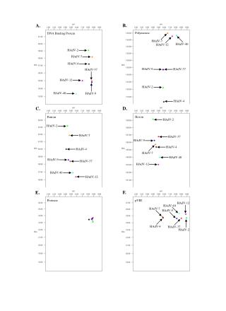
The H2A.Z histone in Drosophila Heterogeneity in nucleosome structure results from incorporation of variant histone proteins into the nucleosome. In contrast to the canonical histones, which are multicopy genes expressed during S-phase of the cell cycle, variant histones are encoded by single copy genes that differ in amino acid sequence from S-phase histones and whose expression is not limited to S-phase. Variant histones allow specialization of nucleosome structure for specific purposes. H2A.F/Z is a family of H2A variants that are highly conserved across species and substantially divergent from S-phase H2A in any given species. H2Av is a variant of H2A.Z in Drosophila. H2A.F/Z typically constitutes 5–10% of total H2A proteins in the chromatin of cells. H2A.F/Z may play a role in transcriptional regulation since in Drosophila its incorporation into chromatin during development is coincident with the start of zygotic gene expression. Leach et al. J. Biol. Chem. 275, 23267 (2000) SS 2009 – lecture 1 Biological Sequence Analysis 23
Leach et al. J. Biol. Chem. 275, 23267 (2000) SS 2009 – lecture 1 Biological Sequence Analysis 24