Download
1 / 24
240 likes | 385 Views
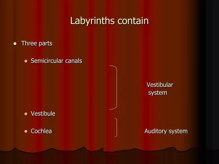
Labyrinths contain. Three parts Semicircular canals Vestibular system Vestibule Cochlea Auditory system. Vestibular system. Function: Balance and equilibrium Sensory cells: Hair cells. Auditory system: Cochlea. Name derived from snail-like shape

E N D
Labyrinths contain • Three parts • Semicircular canals Vestibular system • Vestibule • Cochlea Auditory system
Vestibular system • Function: Balance and equilibrium • Sensory cells: Hair cells
Auditory system: Cochlea • Name derived from snail-like shape • Tube of decreasing diameter coiled around itself • Coiled around a central bony canal called modiolus • Broad base, narrow apex • Length: About 35 mm • In human beings: 2 and 5/8 coils
Isolated cochlear turns from the inner ear of fetal sheep: Photo courtesy of Gerhardt, K.
Cochlear macrostructure • Partially divided throughout its length by a thin spiral shelf of bone called osseous spiral lamina • At the outer (lateral) wall of the cochlea: Spiral ligament
Between the osseous spiral lamina and the spiral ligament: Basilar membrane • Runs all the way along the length of the cochlea, except for a small opening at the apex called the helicotrema • Reissner’s membrane projects diagonally from spiral lamina to outer bony wall of cochlea • Joins the basilar membrane at the helicotrema
Between the osseous spiral lamina and the spiral ligament: Basilar membrane • Runs all the way along the length of the cochlea, except for a small opening at the apex called the helicotrema • Reissner’s membrane projects diagonally from spiral lamina to outer bony wall of cochlea • Joins the basilar membrane at the helicotrema http://www.iurc.montp.inserm.fr/cric/audition/english/ear/fear.htm http://epl.meei.harvard.edu/~hwang/3Dviewer/3Dviewer.html
Cross-section of the cochlea • Basilar membrane and Reissner’s membrane divide the cochlear canal into three ducts: • Scala vestibuli • Scala tympani • Scala media
Scala Vestibuli • Above Reissner’s membrane • Extends from oval window in the vestibule to the helicotrema • Contains perilymph
Scala Tympani • Below the basilar membrane • Extends from round window to the helicotrema • Contains perilymph
Scala media/cochlear sac • Bound below by basilar membrane • Bound above by Reissner’s membrane • Bound on the outer side by Stria vascularis • Contains endolymph
Dimensions of the cochlear partitions • Cochlea: Narrower toward the apex • Basilar membrane: Narrower at the base and wider toward the apical end • Apical end: Flaccid and under no tension • Base end: Stiff and under a small amount of tension
Cochlear microstructure • Organ of Corti • Lies on the basilar membrane in the scala media • Contains many different types of specialized cells
Pillar cells or rods of Corti • Provide structure and support • Inner and outer pillar cells • Form a tunnel called tunnel of Corti • Contains fluid called cortilymph
Hair cells • On the outer side of the outer pillar/rod cells: Outer hair cells (OHC) (Away from modiolus) • On the inner side of the inner pillar/rod cells: Inner hair cells (IHC) (Toward the modiolus)
Supporting cells • OHC supported by cells of Deiter and Hensen • IHC supported by border cells of the inner sulcus
Reticular lamina Formed partly by the phalangeal processes of the Deiter’s cells.
OHC • Around 12000 in number • Test-tube shaped • Three-five rows
IHC • Around 3400 in number • Flask shaped • Single row
Stereocilia • Hairlike projections • Project from the top of IHC and OHC • Graded in length • Have cross-bridges called tip-links
Stereocilia of OHC and IHC • OHC: • 6-7 rows per OHC • Each row has a W shaped arrangement • IHC: • 2-4 rows per IHC • Each row in a shallow U-shaped arrangement
Tectorial membrane • Gelatinous transparent membrane projecting from spiral lamina • Attaches loosely on outer edge to the Deiter’s and Hensen’s cells. • Longest stereocilia of OHC embedded in inferior surface of tectorial membrane • Stereocilia of IHC not embedded in tectorial membrane