Download
1 / 55
661 likes | 1.48k Views
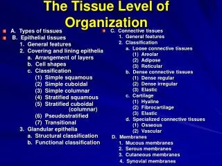
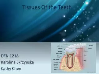
Tissue of the teeth. Dr Jamal Naim PhD in Orthodontics. Periodontium. Introduction. The periodontium is defined as those tissues supporting and investing the tooth and consists of: Cementum Periodontal ligament (PDL) Bone lining the alveolus Parts of the Gingiva facing the tooth

E N D
Tissue of the teeth Dr Jamal Naim PhD in Orthodontics Periodontium
Introduction The periodontium is defined as those tissues supporting and investing the tooth and consists of: • Cementum • Periodontal ligament (PDL) • Bone lining the alveolus • Parts of the Gingiva facing the tooth • All of these tissues has ectomesenchymal origin.
Cementum It is a specialized hard connective tissue that shares some physical, chemical, and structural characteristics with compact bone. Unlike bone, however, human cementum is avascular.
Cementum Cementum covers the anatomic roots of human teeth. It begins at the cervical portion of the tooth at the cemento-enamel junction and continues to the apex. Cementum furnishes a medium for the attachment of collagen fibers that bind the tooth to surrounding structures.
Chemical composition • 50-55% of weight organic material and water. • Collagen fibrils • Protein polysaccharides (proteoglycans) • 45-50% inorganic material. • Hydroxylapatite (calcium & phosphate) • Various trace elements, (Fluor, copper, zinc etc.) • the highest fluoride concentration of all tooth tissue
Cementogenesis • It takes place in two phases: • Matrix formation • Mineralization • There are 3 cell types responsible for the cementogenesis: • Cementoblasts • Cementocytes • Fibroblasts • All of these cells are differentiated ectomesenchymal cells.
Root formation Root development begins after the crown is formed. Root formation precedes by deposition of dentin along the inner aspect of the Hertwig`s root sheath. HERS
Root formation HERS derives from the corono-apical extension of the inner and outer dental epithelium.
Cementogenesis It is believes that HERS induce the formation of the root dentin. Ectomesenchymal cells of the dental pulp differentiate into odontoblasts to form the first layer of root dentin. Just before degeneration of the Hertwig`s root sheath, a thin cell-free layer of cementum is formed on the surface of dentin: intermediate cementum.
Cementogenesis After degeneration of the epithelial root sheath ectomesenchymal cells from the inner portion of the dental follicle differentiate and become cementoblasts.
Cementogenesis Cementoblasts are large cubical cells showing all features characteristic of protein synthesizing cells. They contain a well developed RER, Golgi appartus, numerous mitochondria and a large nucleus. They elaborate the organic matrix which is called cementoid. Cementoid consists of collagen fibers and a ground substance composed of proteoglycans. The cementum is laid down in a rhythmic process (increments) until the full thickness is reached.
Cementogenesis cementoid Cementodentinal junction Cementoblasts
Cementogenesis After reaching the full thickness the cementoblasts enter a quiescent stage. During matrix formation fibroblasts form collagen fibers (sharpey`s fibers), which become embedded in the matrix to provide attachment of the root to the surrounding bone. Those fibers are also called perforating fibers. There is usually a cementoid layer observed on the cemental surface lined by cementoblasts.
Cementogenesis The mineralization begins after forming the first layer of matrix. Ca and Ph ions from the tissue fluid are deposited ito the matrix and arranged as hydroxylapatite crystals parallel to the fibrils. Sometimes crystals can be seen clustered into groups of nucleation centers as found in bone.
Cementogenesis INTERMEDIATE CEMENTUM: Intermediate cementum is a thin, acellular, amorphous layer of hard tissue approximately 10 micron thick. It is deposited by the inner layer of the epithelial cells of the root sheath. Deposition occurs immediately before the epithelial root cells disintegrate as a sheet and migrate away from the root into the periodontal tissue. The remnants of this sheath are called cells of malassez
Cementogenesis intermediate cementum Cementoblasts
Cementogenesis Remnants of the Hertwig`s root sheath, which disintegrate into the PDL are the Malassez cells. intermediate cementum
Types of cementum Two types of cementum are recognized depending on the presence or absence of cells, and they are therefore known as: • acellular cementum • acellular afibrilar cementum (over enamel) • acellular fibrilar (extrinsic fiber) cementum and • cellular fibrilar (intrinsic fiber) cementum Both forms are deposited in layers and the deposition probably continues throughout life.
Types of collagen fibers of C. The collagen fibrils of cementum are of two kinds: • The first group is made up of the embedded parts of the principal fibers of the periodontal Ligament, which are known as sharpey's fibers (extrinsic group). They formed by the fibroblasts of the periodontal ligament. • The other group of collagen fibrils, which constitute the intrinsic group, are formed by the cementoblasts and are found between the sharpey's fibers arranged either randomly or parallel to the surface of the cement.
Acellular cementum Acellular C. consists of calcified intercellular substance and embedded collagen fibers from the extrinsic group (sharpey's fibers). The remainder fibers are from the intrinsic group and run perpendicular to the sharpey's fibers. In dried ground section the sharpey's fibers disintegrate and appear dark. Acellular C. Dentin
Acellular cementum • It predominates in the coronal half of the root. • It may also cover the root from cemento-enamel-junction to the apex, but always the apical third of the root is predominant cellular.
AC CC Dentin Dentin
Acellular Afibrilar cementum A type of cementum formed on the cervical part of the enamel for a short distance. It is formed due to the degeneration of the reduced dental epithelium covering the cervical area of the enamel before eruption.
A fibrilar cementum Enamel Dentin A fibrilar cementum cementum
Cellular cementum During the calcification cementoblasts are incorporated in the cementum. The cells become cementocytes. They lie in spaces designated as lacunae. Atypical cementocyte has numerous cell processes, or canaliculi, radiating from its cell body mainly toward the PDL (the source of nutrition). Cementocytes have less activity than cementoblasts. They are irregularly distributed throughout the thickness of CC.
Cellular cementum Cementocytes PDL Dentin
Cementocytes Dentin Cementodentinal junction Cellular cementum
Cementocytes PDL side Lacunae and canaliculi
Cellular cementum It is a less mineralized type of C., which is formed after tooth is in occlusion. It is more frequent on the apical half o the root. It is always formed on the surface of acellular cementum. It is less in anterior teeth, but thicker in multi-rooted teeth. Sometimes the cellular cementum covers the inner wall of dentin forming a lining of the root canal.
Cellular cementum The cellular cementum has only about 60% of its collagen fibers derived from sharpey's fibers. The remainder fibers are intrinsic fibers.
Structures of cementum • Incremental lines of Salter • Cementodentinal junction • Cementoenamel junction • Sharpey's fibers • Cementicles
Incremental lines of Salter In both acellular and cellular cement incremental lines run roughly parallel with the root surface. These are formed by fiber-free amorphous substance and represent intervals between successive deposition of cement and are called incremental Lines of Salter. Histochemical study indicates that the incremental lines are highly mineralized areas with less collagen and more ground substance.
Incremental lines of Salter Dentin
Cementodentinal junction The dentin surface upon which cementum is deposited is relatively smooth in permanent teeth. The cementodentinal junction in deciduous teeth, however, is sometimes scalloped. Permanent teeth Deciduous teeth
Cementodentinal junction Cementodentinal junction
Cementoenamel junction The relation between, cementum and enamel at the cervical region of teeth is variable. In approximately 30% of all teeth, cementum meets the cervical end of enamel in a relatively sharp line.
Cementoenamel junction In about 10% of the teeth, enamel and cementum do not meet. Presumably this occurs when enamel epithelium in the cervical portion of the root is delayed in its separation from dentin
Cementoenamel junction In such cases these is no cementoenamel junction. Instead, a zone of the root is devoid of cementum and is, for a time, covered by reduced enamel epithelium.
Cementoenamel junction In approximately 60% of the teeth, cementum overlaps the cervical end of enamel for a short distance. This occurs when the enamel epithelium degenerates at its cervical termination, permitting connective tissue to come in direct contact with the enamel surface.
cementum overlaps enamel no cementoenamel junction
Sharpey's Fibers At the surface of the cement the principal fibers of the periodontal Ligament pass into its substance. The fibers, which embedded in the cement, are known as sharpey's fibers. In the acellular cementum sharpey's fibers are usually calcified. In the cellular cementum each fiber commonly shows an uncalcified core with a calcified Periphery. In dried ground sections they appear as dark lines as a result of their disintegration
Sharpey's Fibers Sharpey's fibers always emerge from the cement in a straight line and continue across the periodontal interval and into the alveolar bone stresses are then always applied in the direction of their long axis.
Cementicles Cementicles are small-mineralized bodies, which may be found in the periodontal ligament. They may be attached to the cementum or the alveolar bone, or occur free in the periodontal ligament. When present, cementicles are generally found about all or most of the teeth. Cementicles may be formed by mineralization of degenerating epithelial rests or thrombosed vessels.
to bind the tooth to alveolar bone.
Age changes of cementum Hypercementosis: It is an abnormal thickening of cementum, may be diffuse or circumscribed. It may affect all teeth of the dentition, be confined to a single tooth, or even affect only Parts of one tooth. If the overgrowth improves the functional qualities of the cementum, it is termed a cementum hypertrophy. If the overgrowth occurs in nonfunctional teeth, it is termed hyperplasia (e.g. ostitis deformans paget).
Localized hypercementosis Generalized hypercementosis