Download
1 / 29
290 likes | 505 Views
Bringing light into the chaos: A general introduction to optics and light microscopy. Juliana Schwarz 19/03/07. Overview. Part one: The root of all evil Basic terms and applications in light microscopy. Part two: Bringing colour into the light

E N D
Bringing light into the chaos: A general introduction to optics and light microscopy Juliana Schwarz 19/03/07
Overview Part one: The root of all evil Basic terms and applications in light microscopy Part two: Bringing colour into the light Fluorescence microscopy and special applications: Widefield, Confocal microscopy, Multiphoton, FRET, FLIM,FRAP, Photoactivation, TIRF
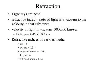
What is light? Light is electromagnetic radiation. What we usually describe as light is only the visible spectrum of this radiation with wavelengths between 400nm and 700nm. The elementary particle that defines light is the photon. b) a) • There are 3 basic dimensions of light • Intensity (amplitude) which is related to the perception of brightness • Frequency (wavelength), perceived as colour • Polarization (angle of vibration) which is not or weakly perceptible to humans
What is a microscope? Theoretically a microscope is an array of two lenses. Focal plane Image plane Image plane Eyepiece lens Objective lens Tube lens Modern microscope with ICS (Infinity Colour corrected System) Classic compound microscope
Your friend - the objective Objectives can be classified into transmitted light and reflected-light (Epi) versions.
Flat-field correction and aberration correction Describe two main criteria for the quality of an objective: Flatness of the intermediate image Elimination of chromatic errors
Spherical aberration Spherical aberration causes beams parallel to but away from the lens axis to be focussed in a slightly different place than beams close to the axis. This manifests itself as a blurring of the image.
Flat-field correction and aberration correction Describe two main criteria for the quality of an objective: Flatness of the intermediate image Elimination of chromatic errors
Chromatic aberration Chromatic aberration is caused by a lens having different refractive indexes for different wavelengths. Since the focal length of a lens is dependent on the refractive index, different wavelengths will be focused on different positions in the focal plane. Chromatic aberration is seen as fringes of colour around the image. It can be minimised by using an achromatic doublet (= achromat) in which two materials with differing dispersion are bonded together to form a single lens.
Objective types elimination of chromatic errors flatness of the intermediate image • CP-Achromat Good colour correction – exactly for two wavelengths. Field flatness in the image center, refocusing also covers the peripheral areas. For fields of view up to dia. 18 mm. Versions for phase contrast. • Achroplan Improved Achromat objectives with good image flatness for fields of view with dia. 20 or even 23 mm. Achroplan for transmitted light and Achroplan Ph for phase contrast. • Plan-Neofluar Excellent colour correction for at least three wavelengths. Field flattening for the field of view with dia. 25 mm. Highly transmitting for UV excitation at 365 nm in fluorescence. All methods possible, special high-quality variants are available for Pol and DIC. • Plan-Apochromat Perfect colour rendition (correction for four wavelengths!). Flawless image flatness for fields of view with dia. 25 mm. Highest numerical apertures for a resolving power at the very limits of the physically possible.
Magnification is defined by the magnification by the objective x the magnification by eyepiece BUT maximum magnification does not mean maximum resolution! What is magnification?
What is resolution? Resolution describes the minimal distance of two points that can be distinguished. Picture taken from http://microscopy.fsu.edu/primer/anatomy/numaperture.html
α2 α1 Oil (n = 1.5) Air (n = 1.0) Coverslip (n = 1.5) Glass slide (n = 1.5) What is the numerical aperture? NA is an estimate of how much light from the sample is collected by the objective Objective lens NA = n sin n = refractive index α = angle of incident illumination
Numerical aperture, NOT magnification determines resolution! Increasing NA A lens with a larger NA will be able to visualize finer details and will also collect more light and give a brighter image than a lens with lower NA.
How can we use the properties of light to create contrast? Which properties can be used? Absorption Scattering Refraction Phase Polarization
Brightfield Phase contrast DIC Darkfield Contrasting techniques Taken from: http://fig.cox.miami.edu/~cmallery/150/Fallsyll.htm
Contrasting techniques • Brightfield • Darkfield • Phase Contrast • Polarization Contrast • Differential Interference Contrast (DIC) • Fluorescence Contrast (Ireen)
Brightfield Principle: Light is transmitted through the sample and absorbed by it. Application: Only useful for specimens that can be contrasted via dyes. Very little contrast in unstained specimens. With a bright background, the human eye requires local intensity fluctuations of at least 10 to 20% to be able to recognize objects. Piece of artificially grown skin (www.igb.fhg.de/.../dt/PI_BioTechnica2001.dt.html ) Cross section of sunflower root (http://www.zum.de/Faecher/Materialien/beck/12/bs12-5.htm) all our microscopes can be used for brightfield
Darkfield Principle: The illuminating rays of light are directed through the sample from the side by putting a dark disk into the condenser that hinders the main light beam to enter the objective. Only light that is scattered by structures in the sample enters the objective. Application: People use it a lot to look at Diatoms and other unstained/colourless specimens Darkfield Symbiotic Diatom colony (www1.tip.nl/~t936927/making.html) Brightfield we do not have microscopes set up for darkfield
Phase ring I0 not aligned aligned Phase stops I Phase contrast in theory Principle: Incident light [Io] is out of phase with transmitted light [I] as it was slowed down while passing through different parts of the sample and when the phases of the light are synchronized by an interference lens, a new image with greater contrast is seen. most of our microscopes are set up for phase contrast
Phase contrast in practice Application: Phase contrast is the most commonly used contrasting technique in this institute. All tissue culture microscopes and the time-lapse microscopes are set up for phase. BUT: MOST OF YOU ARE USING IT IN THE WRONG WAY!! Because you do not use the right phase stop with the corresponding objective! wrong phase stop right phase stop brightfield
Brightfield Polarization contrast Polarization contrast with Lambda plate Polarization Contrast Principle: Polarized light is used for illumination. Only when the vibration direction of the polarized light is altered by a sample placed into the light path, light can pass through the analyzer. The sample appears light against a black background. A lambda plate can be used to convert this contrast into colours. Application: Polarization contrast is used to look at materials with birefringent properties, in which the refractive index depends on the vibration direction of the incident light, e.g. crystals or polymers. Analyzer Lambda plate Polarizer we do not have microscopes set up for polarization contrast
Δ ~Δn/Δx Δ > 0 C n1 A B n2 n3 X ΔX ΔX ΔX D(ifferential) I(nterference) C(ontrast) Principle: Also known as Nomarski microscopy. Uses polarized light for illumination. Synchronizing of the different phases of incident and transmitted light is done by a set of prisms and filters introduced into the light path. most of our microscopes are set up for DIC
Contrasting techniques - a summary • Brightfield -absorption Light is transmitted through the sample. Only useful for specimens that can be contrasted via dyes. Very little contrast in unstained specimens. • Darkfield -scattering The illuminating rays of light are directed from the side so that only scattered light enters the microscope lenses, consequently the cell appears as an illuminated object against the view. • Phase Contrast- phase interference Incident light [Io] is out of phase with transmitted light [I] and when the phases of the light are synchronized by an interference lens, a new image with greater contrast is seen • Polarization Contrast -polarization Uses polarized light for illumination. Only when the vibration direction of the polarized light is altered by a sample placed into the light path, light can pass through the analyzer. The sample appears light against a black background. • Differential Interference Contrast (DIC) – polarization + phase interference Also known as Nomarski microscopy. Synchronizing of the different phases of incident and transmitted light is done by a set of special condenser lens mounted below the stage of a microscope • Fluorescence Contrast (->Ireen)
10x 20x 40x 60x Objective Coverslip-types: 1: 0.13 - 0.17 mm 1.5: 0.16 - 0.19 mm 2.0: 0.19 - 0.23 mm Final words from your friend - the objective
Useful links • Margaret and Tom (ext. 6872)!!! • Zeiss – Microscopy from the very beginning http://www.zeiss.de/C1256B5E0047FF3F?Open • Molecular Expressions homepage http://micro.magnet.fsu.edu/index.html • Wikipedia http://en.wikipedia.org/wiki/Main_Page