Download
1 / 11
110 likes | 119 Views
Know the 2023 latest updates on Age Related Macular Degeneration Treatment, Symptoms to save your eyes! Also, know diabetic macular edema treatment and causes.

E N D
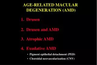
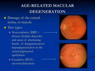
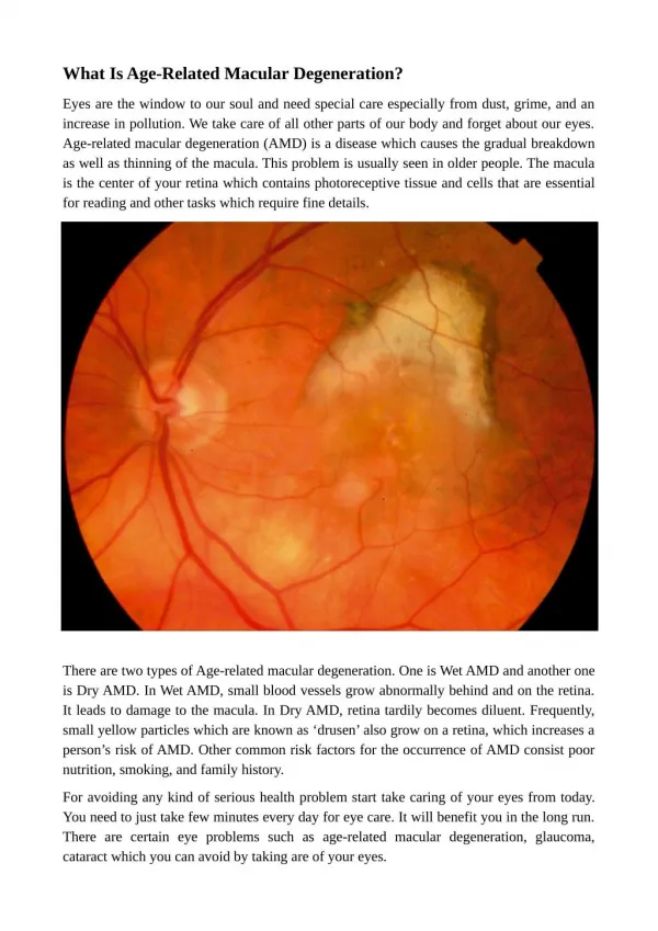
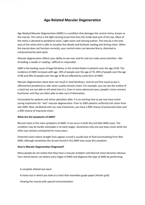
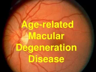
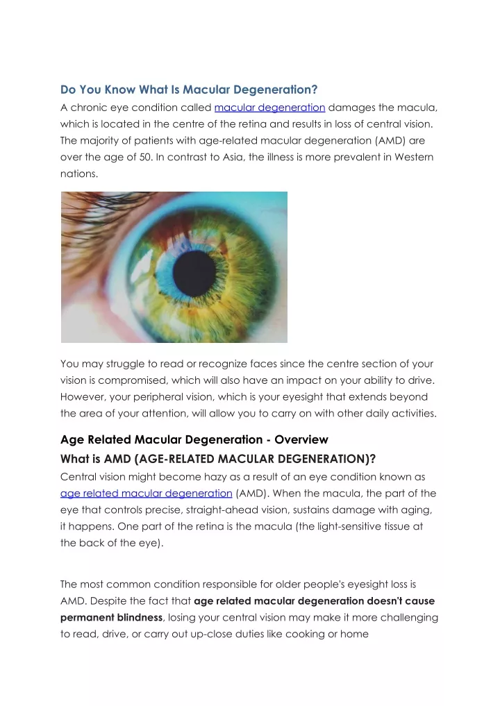
Do You Know What Is Macular Degeneration? A chronic eye condition called macular degeneration damages the macula, which is located in the centre of the retina and results in loss of central vision. The majority of patients with age-related macular degeneration (AMD) are over the age of 50. In contrast to Asia, the illness is more prevalent in Western nations. You may struggle to read or recognize faces since the centre section of your vision is compromised, which will also have an impact on your ability to drive. However, your peripheral vision, which is your eyesight that extends beyond the area of your attention, will allow you to carry on with other daily activities. Age Related Macular Degeneration - Overview What is AMD (AGE-RELATED MACULAR DEGENERATION)? Central vision might become hazy as a result of an eye condition known as age related macular degeneration (AMD). When the macula, the part of the eye that controls precise, straight-ahead vision, sustains damage with aging, it happens. One part of the retina is the macula (the light-sensitive tissue at the back of the eye). The most common condition responsible for older people's eyesight loss is AMD. Despite the fact that age related macular degeneration doesn't cause permanent blindness, losing your central vision may make it more challenging to read, drive, or carry out up-close duties like cooking or home
maintenance. Some people develop AMD relatively slowly, whereas others develop it more quickly. You could take a while to notice visual loss if you have early AMD. To determine if you have AMD, it is crucial to have routine eye exams. What are the different forms and phases of AMD? Dry and moist age related macular degeneration are the two different subtypes. The majority of AMD patients have dry AMD (also called atrophic AMD). At this age, the macula starts to deteriorate. Dry AMD develops in stages that are early, middle, and late. It typically takes years to grow gradually. Although there is no known treatment for late dry AMD, there are ways to make the most of your remaining vision. If you only have one eye with late dry AMD, you can still take measures to protect the other eye. A less common type of late AMD called wet AMD often causes a sudden loss of vision (also known as advanced neovascular AMD). However, wet AMD is always in the late stage and might arise at any stage of dry AMD. The development of abnormal blood vessels behind the eye damages the macula. The good news is that wet AMD treatments are readily available. What AMD symptoms are present? The symptoms of age related macular degeneration change as it progresses. Dry AMD develops in stages that are early, middle, and late. AMD is a degenerative disease, therefore symptoms usually get worse over time. Early-stage dry AMD has no symptoms * Some people with intermediate dry AMD don't experience any symptoms. Others may only experience minor symptoms, such as a slight blurring of their central vision or trouble seeing in low light. * A warning sign of advanced AMD is when straight lines seem wavelike. Visit your eye doctor as soon as possible if you experience this symptom.
What can I do to lower my chances of getting AMD? Making these healthy decisions may help you reduce your risk of AMD (or slow vision loss from AMD), according to research: • Stop smoking or avoid starting. • Engage in regular exercise • Maintain normal levels of cholesterol and blood pressure. • Consume healthful foods, such as fish and leafy greens. My eye doctor's method for detecting AMD AMD can be examined by eye professionals as part of a thorough dilated eye exam. Your doctor will give you some eye drops to expand your pupil before doing a quick and painless eye exam to screen for AMD and other eye issues. Additionally, your doctor might advise having an examination called optical coherence tomography (OCT). Your eye doctor will use a special device to take photos of the interior of your eye during an OCT exam. What is the treatment for Age-Related Macular Degeneration (AMD)? The stage and kind of AMD will naturally affect the course of treatment. Early AMD has no known cure, so your ophthalmologist will likely merely do routine eye exams to monitor your eyes' health. Healthy eating, consistent exercise,
and giving up smoking can all be beneficial. Special dietary supplements (vitamins and minerals) may be able to prevent late AMD from developing if you have intermediate AMD in 1 or both of your eyes. These supplements may decrease the progression of AMD in your other eye if you just have late AMD in one eye. Other therapies may be able to stop additional vision loss if you have wet AMD: * Anti-VEGF medications * Injections and laser therapy are used in photodynamic therapy (PDT). * There is presently no cure for late dry AMD, but doctors are working hard to find a solution. * Additionally, you can seek assistance to help you cope with AMD-related visual loss. How can I manage AMD-related visual loss? Not all AMD patients experience late-onset or bilateral occurrence. Living with AMD-related visual loss, however, can be difficult. Low vision makes it difficult to carry out daily chores, even with the aid of glasses, contact lenses, medication, or surgery The good news is that resources are available, including low-vision equipment and services for vision rehabilitation. With limited vision, vision rehabilitation can teach you the skills you need to maintain your
independence and keep active. Immunity-Boosting Techniques An immune system component known as the "complement cascade" has been directly linked to AMD. The immune system's complement component is thought to target the retina. The retinas of mice can be protected by inhibiting the complement cascade. While two phase II trials by Genentech targeting the complement protein known as factor D in individuals with geographic atrophy were unsuccessful, promising outcomes were seen in a phase II study by Apellis targeting a different complement protein known as C3. Phase III studies for the medication Apellis have begun. Similar results were shown with another medication named Zimura, which inhibits the complement protein C5 and has advanced to a phase II trial. Zimura slows the progression of geographic atrophy. In the middle of the eye's vitreous jelly, both of these medications are injected. Another strategy is to use the oral antibiotic doxycycline to attempt and block particular immune cells. Clinical trials are in phase II for this. Phase II clinical trials for GA are testing an oral medication called ALK-001 as a way to reduce the toxic consequences of vision cells. This medicine is a modified (deuterated) version of vitamin A that prevents the development of the hazardous byproduct A2E. Limiting the workload of the photoreceptors (vision cells) is an alternative strategy that will reduce any possibly hazardous byproducts of that labour.
Emixustat, an oral medication, works to accomplish this by only slightly impairing photoreceptors' capacity to detect light. Some degree of night blindness or trouble seeing in low light is a potential side effect. Sadly, a clinical investigation of patients who had geographic atrophy revealed that this medication did not slow the atrophy's progression. Genetic Therapy Current mouse gene therapy studies show long-term antioxidant or anti-cell death effects. Children with genetic blindness have benefited from clinical trials including retinal gene therapy. Managing blood lipids Due to the involvement of lipid (fat) buildup, AMD is comparable to atherosclerosis, or the hardening of the arteries, in several aspects. Current studies are examining the potential benefits of lipid-controlling medications. What is Diabetic Macular Edema? Diabetes has a condition called diabetic macular edema (DME). DME can occur in people with type 1 or type 2 diabetes.
DME happens when too much fluid begins to accumulate in the macula of the eye. We can focus and see minute details thanks to the macula. It is situated in the middle of the retina, the blood vessel-filled lining in the back of the eye. Vision issues result from the macula's overflowing fluid. DME typically worsens over time. The blood vessels in the retina might become damaged by high blood sugar levels. Fluid leakage from damaged blood vessels can result in edema and other problems. Retinopathy is the term for this injury. Diabetic Macular Edema Types The degree of edema in the retina is frequently used to categorize diabetic macular edema. More edema typically results from a bigger retina, which causes more vision loss. The location of the blood vessel injury may also serve to define it. It can sometimes only occur in a certain location. Other times, the retinal tissue is more severely damaged. Your eye doctor may do a number of tests on your eyes during an eye exam. The tests evaluate any visual loss and reveal any blood vessel damage or the degree of fluid accumulation (swelling) in the retina. The following are typical eye exams to detect DME or evaluate eye damage: CT imaging with optical coherence (OCT). This examination measures any retinal enlargement.
Fundus photography The retina is photographed in great detail during this examination to look for atypical blood vessels. Angiography using fluorescein. A dye is injected into your arm or hand during this examination to make the blood flow in your retina more visible. You'll receive eye drops to dilate your pupils before every test (called pupil dilation). Your eye doctor can now see more of the retina as a result. You won't experience any discomfort while the test is being done, save from minor light sensitivity brought on by the pupil dilation. Early detection and monitoring by an eye doctor can help avoid additional vision loss. Even lost vision may be restored with treatment. In just a few months if untreated, vision can severely deteriorate. DME Signs and Symptoms In the initial stages, there may be no symptoms. If you have diabetes, you should see an eye doctor once a year so that they may inspect your eyes for any changes. Early detection and treatment of retinopathy and diabetic macular edema can prevent or restore visual loss. If you have any of the following symptoms, please notify your eye doctor: • vision blur • seeing washed-out hues • seeing additional floaters in your vision • dual vision DME Causes High blood sugar levels can cause damage to small blood vessels in the eyes over time, increasing the risk of DME. Working with your healthcare team to keep your blood sugar levels as close to target as feasible is critical to maintaining the health of your eyes. Blood vessel damage can also be caused by excessive blood pressure and cholesterol levels.
Pregnancy can increase the chance of getting DME in some forms of diabetes. During pregnancy, your doctor may advise you to get more frequent eye tests. Risk elements There are additional risk factors that might contribute to DME in patients with type 1 or type 2 diabetes. These risk factors are as follows: • inadequate blood sugar control • elevated cholesterol levels • elevated blood pressure • kidney disorder (nephropathy) • Pregnancy sleep apnea Preventing DME It is never too late to consult with your doctor about treatment choices. If you've been diagnosed with DME, initiating treatment as soon as possible will help prevent long-term eye damage and vision loss. When it comes to protecting your vision, taking preventive measures can make a major impact. You may help your eyes by doing the following: • Annual eye exams should be scheduled with your eye doctor. • Contact your eye doctor as soon as you notice any changes in your vision. • Work with your diabetes care team to effectively manage your blood sugar levels. • Take action to maintain healthy blood pressure and cholesterol level. • Inform your healthcare team if you're having trouble controlling your blood sugar. They may advise you to make lifestyle changes, take medication, or take other efforts to keep your blood sugar levels within a safe range. Diabetic Macular Edema Treatment Laser treatment for macular edema treatment * This macular edema treatment therapy is typically provided in a clinical setting, such as your eye doctor's office. * Laser therapy targets damaged parts of the retina with small lasers. This procedure plugs blood vessels that are leaking and stops aberrant blood vessel growth.
* Laser therapy can help you keep your existing vision and prevent further vision loss. * To restore eye damage, you will most likely require numerous laser treatments over time. If more eye damage occurs, you may require additional therapy. Injectable medications Injectable drugs are classified into two types: anti-VEGF and steroids. There are various types accessible within each group. Your eye doctor will establish the appropriate medicine and frequency of treatment for you. The drug is injected into your eye by your eye doctor using a very thin needle. When doctors provide this drug, they will numb your eye to prevent pain. Anti-VEGF is an acronym that stands for "anti-vascular endothelial growth factor." Medications in this category help to prevent aberrant blood vessel formation, which could lead to further eye damage. They also help to minimise edema. In general, anti-VEGF medications: have a high success rate in improving eyesight, help lower the quantity of fluid that seeps into the retina, have a minimal risk of consequences, and are deemed safe. Anti-VEGF injections are typically not unpleasant. If needles make you nervous, talk to your doctor about options for keeping you calm throughout the treatment. Steroids are another option for macular edema treatment. Steroids may be used if anti-VEGF drugs no longer function to help reduce retinal edema and improve vision.
In some circumstances, however, steroids may raise the risk of cataracts. Your doctor will determine whether the benefit of this therapy outweighs the danger. Steroid treatment for DME may be provided in the form of single injections or implants that gradually release the medicine. Summary: Macular degeneration is a chronic eye condition that causes loss of central vision because of damage to the macula in the central part of the retina. There is a lot of research being done on age-related macular degeneration (AMD), which promises better therapies.