Download
1 / 16
160 likes | 400 Views
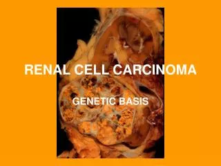
6- Renal Carcinoma . Renal cell carcinoma occupying the lower renal pole . Cross-section of kidney shows a well circumscribed yellowish tumor mass occupying lower pole of the kidney. There is compression of the hilar blood vessels and renal pelvis (small arrow). .

E N D
Renal cell carcinoma occupying the lower renal pole Cross-section of kidney shows a well circumscribed yellowish tumor mass occupying lower pole of the kidney. There is compression of the hilar blood vessels and renal pelvis (small arrow).
Section shows clear tumor cells with mild pleomorphic nuclei
Clear cell carcinoma of the kidney:Section of the kidney shows: • Compressed kidney tissue at the margin of the tumor masses. • Tumor cells are large polygonal with clear cytoplasm (dissolved glycogen and lipid) and pyknoticnuclei. • Cells are arranged as alveolar groups or tubules or papillary formations separated by thin fibro-vascular septae. • Cells show some pleomorphismand mitosis. • Areas of haemorrhage and necrosis can be seen.
Wilms Tumor Gross picture shows partly pale and partly hemorrhagic solid tumour replacing almost the entire renal parenchyma and areas of necrosis also seen .
Wilm's tumor resembles fetal nephrogenic zone of the kidney. • 3 major components: • Undifferentiated blastema cells , • Epithelial tissue attempts to form primitive glomerular &tubular structure • Mesenchymal (stromal) tissue
8- Carcinoma of the urinary bladder
Longitudinal section of urinary bladder and prostate showing • benign prostatic hyperplasia , • trabeculation of the urinary bladder wall and • bladder carcinoma (asterix )which is most likely proved histologically to be Transitional cell carcinoma .
[UROTHELIAL CARCINOMA OF THE BLADER ; LOW GRADE]. Multiple papillary projections lined by multiple layers of urothelium (transitional epithelium). These low grade tumors show overall preservation of cell polarity, few mitoses, and lack of significant morphologic atypia.
Papillary Urothelial Carcinoma, High-grade The nuclei in this high-grade tumor are significantly enlarged and show variably increased chromatin content