Download
1 / 53
570 likes | 1.58k Views
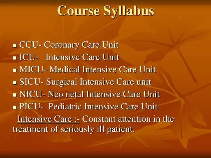
Course Syllabus. CCU- Coronary Care Unit ICU- Intensive Care Unit MICU- Medical Intensive Care Unit SICU- Surgical Intensive Care unit NICU- Neo netal Intensive Care Unit PICU- Pediatric Intensive Care Unit

E N D
Course Syllabus CCU- Coronary Care Unit ICU- Intensive Care Unit MICU- Medical Intensive Care Unit SICU- Surgical Intensive Care unit NICU- Neo netal Intensive Care Unit PICU- Pediatric Intensive Care Unit Intensive Care :- Constant attention in the treatment of seriously ill patient.
Patient Assessment Vital Sign BP- Blood pressure 120/84 m.m Hg T.P.R Temperature- Pulse Respiration 98.4 ˚ F 70 to 80/mint 18/Mint
Eupnea (Normal) • Dyspnea – Difficulty in breathing • Apnea- Absence of Breathing • Orthopnea- Discomfort of Breathing- That is aggravated by lying down. • Tachypnea • Bradypnea- • Cheyne stokes
Hypoventilation: Reduced alveolar ventilation relative to metabolic carbon- Dioxide production so that alveolar carbon- dioxide Pressure increases above normal (under ventilation) • Hyperventilation: Increase alveolar ventilation relative to metabolic carbon-dioxide production, so that alveolar Carbon-dioxide Pressure decreases to below normal. (Over ventilation)
6. Sleep apnea- Cessation of breathing during sleep 7. Cyanosis- Dark bluish discoloration of the skin and mucus membrane due to deficient oxygenation of the blood. 8. Hypoxia- Decrease below normal level of oxygen in inspired gases, arterial blood or tissue without reaching anoxia. 9. Hypoxemia- Sub normal oxygenation of arterial Blood Short of Anoxia.
10. Anoxia - Absence or almost complete absence of oxygen from inspired gases, arterial blood or tissue 11. Hyper Capnia – Abnormally increased arterial carbon dioxide tension. 12. Hypocapnia –Abnormal decreased arterial Carbon dioxide tension. 13. Asphyxia- Impaired or absent exchange of oxygen and carbon dioxide on a ventilatory basis combined hypercapnia and hypoxia or anoxia. 14. Tachycardia – Rapid beating of the heart.
Conventionally applied to rates over 90 beates per minute. Causes. Sinus tachycardia. Axiety-exercise-pain-fever-sepsis-hypovolaemia-heart failure-pulmonery embolism-pregnancy-thyrotoxicosis-beriberi-co2 Retention-Autonomicneuropathy-sympathomimatice.g caffeine, adrenline.
15.Bradycardia Slowness of the heart beatusually defined (by convention) as rate under 60 beates per minute. Causes Physical fitness-vasovagal attacks-sick sinus syndrome-acute- hypothyroidism-hypothermia-increase intracranial pressure-cholestasis.
16. Hypotention. Subnormal arterial blood pressure. 17. hypertention. High blood pressure. Transitory or sustained elevation. systolic blood pressure above 140 mm Hg.diastolic blood pressure above 90% mm Hg.Primary cause. Unknown 95% of cases.Secondary causes. Hypertention 5% cases causes include, Renal diseases, Endocrine diseases.
18.palpitation • Strong or irregular heart beats pulsation of the heart, perceptible to the patient, usually with an increase in frequency or force with or without irregularity in rhythm.
Critical disorders • Arrythmias- distrubance of cardiac rhythem • CVA- cerebro vascular accident. • M.I- myocardiac infarction. • C.C.F- congestive cardiac failure • I.H.D- ischemic heart disease • A.Fib- atrialfibrilation • V.Fib- ventricular fibrilation • ARDS- Acute Respiratory Distress Syndrome. • Pulmonary embolism:- Obstruction or occlusion of the pulmonary artery. • Pulmonary edema:- An accumulation of an excessive amount of watery fluid in cell or intercellular tissues. • Trauma- an injury physical or mental
CRITICAL DISORDERS • Arrhythmias • Cardiac arrhythmia, also called dysrhythmia, is an irregularity or the loss of normal rhythem of the heart beat. • Cerebro Vascular Accident (CVA) • A stroke, also known as a cerebro vascular accident or CVA, is damage to the brain that occurs when the blood flow to the brain is disrupted because a blood Vessel Supplying it is either blocked or has ruptured. • Myocardial Infarction (MI) • Also known as a heart attack or MI, is the occlusion (Closing off) of a coronary artery resulting in an infarct of the affected myocardium, Damage to the myocardium impairs the heart’s ability to pump blood through the body.
Congestive Cardiac Failure(CCF) • OR • Congestive Heart Failure (CHF) • Is a syndrome in which the heart is unable to pump enough blood to meet the body’s needs for oxygen and nutrients. In response to the reduced blood flow, the kidneys retain more fluids with in the body and this fluid accumulates in the legs, ankles and lungs. The term congestive refers to this fluid buildup. • Ischemic Heart Disease (IHD) • Also known as IHD is a group of Cardiac disabilities resulting from an insufficient supply of oxygenated blood to the heart that is usually associated with Coronary Artery Disease (CAD). • Fibrillation • Is rapid, random and ineffective Contractions of the heart.
Atrial Fibrillation (A Fib) • Also known as A. Fib the atria beat faster than the Ventricles. This produces an irregular quivering action of the atria and a very rapid ventricular heart beat. • Ventricular Fibrillation (V Fib) • Also known as V. Fib is the result of irregular contractions of the Ventricles and is fatal unless reversed by electric defibrillation. • Acute Respiratory Distress Syndrome (ARDS) • Also known as ARDS, is a type of lung failure resulting from many different disorders that cause pulmonary edema. There are many causes of ARDS including severe infection, Shock, Pneumonia, Burns and Injuries.
Pulmonary Embolism • Obstruction or occlusion of the pulmonary artery. • Pulmonary Edema • Is an accumulation of an excessive amount of watery fluid in cell or Intercellular tissues. • Trauma • An injury physical or mental.
Diagnostics • ECG- Electro Cardiogram. • CXR- Chest X-ray. • ABGS- Arterial blood gas. • PFT- Pulmonary Function Test. • Bronchoscopy- is the examination of the respiratory apparatus with a flexible bronchoscope for diagnostic or treatment purposes. The bronchoscope is introduced nasally and slowly lead down the trachea until the desired level is reached. • Laryngoscopy- Inspection of the larynx by means of the laryngo scope.S • Endoscopy- esophago-gastro-dnodenoscopy- is the examinition of the oesophagus, stomach and upper intestine using a gastroscope. • ECHO- Echocardiography- the use of ultra sound in the investigation of the heart and great vessels and diagnosis of cardio-vascular lesions. • C.T scan- computed tomography. • E.T.T- Exercise tolerence test. • Holter monitor- a technique for long term , continuous usually ambulatory recording of electro-cardiographic signals on magnetic tape for scanning and selection of significant but fleeting changes that might otherwise escape notice. • Angio-graphy- X-Ray imaging of the heart and great vessels made visible by injection of a radio-opaque solution.
DIAGNOSTIC PROCEDURES OF THE CARDIOVASCULAR SYSTEM • Radiology • In conventional radiology also known as X-Ray or Radiography, an image of hard tissue internal structures is created by the exposure of sensitized film to x-radiation (Radio means Radiation and Graphy means the process of Recording). The resulting film is known as an X-Ray or Radiograph (Radio means Radiation and Graph means the resulting Record). Radiographs are made up of shades of gray. • Radiopaque - Hard tissues such as bone and tooth enamel, do not permit x-rays to pass through and they appear white or light gray on the radiograph. • Radiolucent - Air and soft tissues do permit x-rays to pass through and they appear as shades of gray to black on the radiograph.
Projections: • Projections PA - Posterio-Anterior the x-rays travel from posterior to anterior in PA View you can see Enlarge heart pericardial effusion CCF, pulmonary Edema pleural effusion. • Projection AP - Anterio-posterior the x-rays travel from Anterior to posterior. Mostly AP view taken in the ICU or CCU for Heart and Lungs for Ambulant patient. • Projection Lateral - Left lateral view for left Atrium. Enlargement. • Projection Oblique - Lateral or oblique Projections may be useful in detecting Aortic or mitral valve calcification which may be obscured by the spine on the P.A View. How ever echocardiography is more sensitive
ECG • An electrocardiogram also known as ECG or EKG, is a record of the electrical activity of the myocardium it is the process of recording this activity. Electro means Electric, Cardio means Heart and Graphy means the process of recording. In ECG you can see. • Atrial Ventricular Hypertrophy • Myocardial ischaemia and Infarction • Arrhythmias • Effect of cardiac drugs especially digitalis B-Blocker. • Disturbances in electrolyte metabolism specially potassium
ETT (Exercise Tolerance Test) • Stress tests are ECG’s used to asses cardiovascular health and function during and after the application of stress such as exercise on a Treadmill. • Holter Monitor • Use electrodes attached similarly to an ECG but is worn by the patient to record heart rates and rhythem over a 24-hour period. • Echo Cardiography • Also known as an ECHO, is an ultrasonic diagnostic procedure used to evaluate the structures and motion of the heart (Echo means Sound, Cardio means Heart and Graphy means to Record). The resulting record is an echocardiogram. • Assessment of left ventricular function common indications for Echo cardiography. • Diagnosis and Quantification of severity of valve disease. • Identification of vegetations in endocardits. • Detection of pericardial effusion.
Trans esophageal echocardio graphy • Also known as TEE, is a ultrasonic procedure that images the heart from inside the oesophagus. Because the esophagus is so close to the heart, this technique produces clearer images than those obtained with echocardiography in Endocarditis patient with prosthetic (Specially mitral) valve dysfunction, congenital abnormalities (e.g Atrial septal defact). Aortic dissection, infective endocarditis and systemic embolism. • Angiography • Is a radiographic (X-Ray) study of blood vessels after the injection of contrast medium the resulting film is an angiogram this procedure is frequently performed in conjunction with cardiac catheterization.
Angiocardiography • Uses a contrast medium and chest X-Rays to visualize the dimensions of the heart and large blood vessels (Angio means Blood Vessel, Cardio means Heart and Graphy means the process of recording). The contrast medium, which appears while on the film, is used to make these soft tissue structures visible on the resulting angio cardiogram. • Cardiac Catheterization • Also known as CC, is a procedure in which a Catheter is passed into a vein or artery and is guided into the heart. When the Catheter is in place a contrast medium is introduced to produce angiograms to determine how well the heart is working. This procedure is also used for Treatment purposes. Balloon angioplasty and stenting.
Biopsy – It is a diagnostic method in which tissues are removed from the body for examination under microscope. • Pleural Biopsy – Tissues are removed from the pleura for examination under microscope.
Pathology of the Cardiovascular System Coronary Artery Disease Coronary Artery Disease, also known as CAD, is atherosclerosis of the coronary arteries that may cause angina pectoris, myocardial infarction, and sudden death. End-stage coronary artery diseaseis the final phase of CAD characterized by unrelenting angina pain and a severely limited lifestyle in which medication is the only remaining treatment option. Atheroscelerosis (ath-er-oh-skleh-ROH-sis)ishardening and narrowing of the arteries due to a buildup of cholestrol plaques (athero means plaque or fatty substance and sclerosis means abnormal hardening ).
An atheroma (ath-er-OH-mah), which is characteristic of atherosclerosis, is a plaque (fatty deposit) within the arterial wall (ather means plaque and oma means tumour). • This type plaque (PLACK) is similar to the buildup of rust inside a pipe and it may protrude outward into the opening of the vessel or move inward into the wall of the vessel. • Ischemia (iss-KEE-mee-ah) is a deficiency in blood supply due to either the constriction or the obstruction of a blood vessel (isch means to hold back and emia means blood) • Ischemic heart disease (iss-KEE-mick), also known as IHD, is a group of cardiac disabilities resulting from an insufficient supply of oxygenated blood to the heart that is usually associated with CAD. • Angina pectoris (an-JIGH-nah or AN-jih-nuh PECK-toh-riss) is severe episodes of spasmodic choking or suffocating chest pain. This is usually due to interference with, but not complete blockage of, the supply of oxygen to the myocardium. • A myocardial infarction (my-oh-KAR-dee-al in-FARK-shun), also known as a heart attack or MI, is the occlusion (closing off) of a coronary artery resulting in an infarct of the affected myocardium. Damage to the myocardium impairs the heart’s ability to pump blood through the body. • An infarct (IN-farkt) is a localized area of necrosis (tissue death) caused by an interruption of the blood supply.
Moderate myocardial ischemia ! Angina pectoris Moderate atherosclerotic narrowing of lumen Severe acute myocardial ischemia and infarction Occlusion of left coronary artery A myocardial infarction ("heart attack") on left side of the heart Complete/almost complete occlusion, with hardening due to calcium deposition
Emergency Actions • CPR- Cardio – Pulmonary resuscitation • BCLs:- basic cardiac life support • ACLs. Advance cardiac life support • E.T.T Intubation. Endo Tracheal tube intubation. • Defibrilation. The arrest of fibrillation of the cardiac muscle (atrial or ventricular) with restoration of the normal rhythem if successful.
Oxygenation- Addition of oxygen to any chemical or physical system. 1. Specifically used to describe interventions that provide greater oxygen supply to lungs and thus the circulation. • Nebulization- Spraying – vaporization • Monitoring- • Performance and analysis routine measurement aimed at detecting a change in the environment or health status of population
2. On going measurement of performance of a health service. 3. Continuous over sight of implementation of an activity.
CARDIO PULMONARY RESUSCITAION • Emergency life support involves a variety of methods and procedures designed to deal with sudden, life threatening events caused by cardiac or respiratory failure. • Emergency life support traditionally consists of two related phases: • Basic life support or BLS, and • Advanced cardiac life support or ACLS. • Basic life support aims either to
prevent cardiac or respiratory arrest through prompt identification and Inter vention or • Support failed circulation and respiration via cardio pulmonary resuscitation (CPR) • Basic life support should be started by any person present at the time the Incident occurs. Trained Individuals should begin CPR as soon as possible and local emergency medical system (EMS) must be activated mmediately. • Advance cardiac life support includes the essential elements of BLS but provides additional measures not used by lay personal.
These measures includes • Supporting oxygenation • Ventilation with Adjunct equipment • Starting an I \ V route, administering drugs, • Monitoring Cardiac function • Controlling arrhythmias and • Providing post resuscitative care. • While BLS can be initiated and conducted by lay personnel, ACLS requires, physician supervision, either in person or by way of Telecommunication.
Determination the Need for Emergency Life Support. • The primary Indication for emergency life support is Respiratory and Cardiac arrest. • The most common causes of Respiratory arrest are. • Drug over dose • Drowning • Suffocation • Stroke • Electrocution • Smoke Inhalation • Acute airway obstruction by a foreign body. • Airway obstruction due to In consciousness or Coma • Like respiratory arrest, Cardiac arrest has many potential causes. • The most frequent causesof cardiac arrest are. • Electrical disturbances • or arrhythmias • associated with Myocardial Ischemia • or acute myocardial Infarction.
If respiratory arrest occurs first the heart will normallycontinue to beat for a few minutes. Such patients frequently have a pulse. How ever without Ventilation, body storesof oxygen are rapidly used up. The resulting tissue hypoxia quickly impairsbothCardiac and neural function. • How everrapid recognitionandtreatmentofrespiratory arrest can preventa secondary Cardiac arrestand decrease the likelihood ofpermanent brain damage. • If on the other hand, theprimaryevent iscardiac arrest, no oxygen is circulated and tissue hypoxia occurs almost immediately. • A secondary respiratory arrest quickly follows asthe brain stem is deprived of its oxygen supply. • When Ventilation and Circulation both cease, a condition of clinical death exists. With early Treatment clinical deathisreversible.How ever shouldtissue hypoxiabe prolonged,irreversible cell damage occursresultinginbiological death.
Exactly how much time can elapse before clinical deathbecomebiological deathisunknown. Generally biological death occursfourtosix minutes after ventilation and circulation cease. How evermany factorsmay extend this time period. IncludingHypothermia and certain drugs. • In fact some organs can Sustain Periods of anoxia of 15 minutes or more. The primary exception is the brain. • Although other organs may be successfully resuscitated after prolonged periods of anoxia, brain deathmay occurmuch more rapidly,resulting inirreversible loss of cerebral function. • Thus, thegoal of emergency life supportis toreverse clinical deathbyrestoring ventilation and circulation before brain deathcanoccur. Emergency life support should be initiated when ever thesigns of clinical deathare recognized and continue until the patient is declareddead by a physician. Since firm evidence regarding brain death israrely availableduringresuscitation,the decision tostop emergency life supportis as much amoralandethical Judgmentas aclinical one.
BASIC LIFE SUPPORT (BLS) • Basic life support aims to restore ventilation and circulation to victims ofairwayobstruction, and respiratoryand cardiacarrest, all without using equipment. These skills can be used by a single practitioner to restore Ventilation and circulation until the victim is revived or until advance cardiac life support (ACLS) equipment and personnel are available. • In sequence the steps in BLS are • Initial assessment to determine unresponsiveness • Activation of the emergency medical system (EMS) • Airway restoration • Ventilation Breathing • Restoration of circulation • The last three steps are the Traditional ABC’s of resuscitation • A) irway • B) reathing • C) irculation
Each of the ABC’s start with an assessment phase. • The First phase determines unresponsiveness • The Second determines breathlessness. And • The Third determines pulselessness. • Determining Unresponsiveness • Basic life support begins when an otherwise conscious victims is found in an unresponsive or collapsedstate. Since many hospitalized patients exhibit decreased level of consciousness, practitioners can avoid needless intervention by being fully awareoftheir patient’s mental status. • When coming upon a collapsed victim out side the hospital setting who appears unconscious you shouldfirst lookfor any obvioushead or neckinjuries .If suchinjuries are apparent, great care should be taken in subsequentmanipulationof theneckand in anyefforttomovetheindividual. • What ever the location you quickly assess the individual’s level of consciousness by Tapping or gently shaking the shoulder and shouting “Are you ok? If this fails stir (move) the victim, the practitioner should call for help and activate the emergency medical system.(EMS)
Restoring the Air way • Head Tilt / chin lift • Jaw Thrust maneuver • Restoring ventilation (Breathing) • Mouth to Mouth Ventilation • Mouth to Nose Ventilation • Restoring circulation • Pulse – Carotid (Adult) • Brachial or Femoral (Infants) • Cardio pulmonary resuscitation • Although respiratory and cardiac arrest may occur separately, most often they happen to together. For this reason you will usually combineartificial ventilation with cardiac compression. The two combined together are aptly called cardiopulmonary resuscitation or CPR can be ad ministered by one person alone or by Team of Tow or more individuals.
MANAGEMENT • O2 Via nasal cannla (N.C) • Mask Oxygen can be given by mean of mask Disposable polythene mask is light, handy and cheaper • ventury mask (VMK) A Supplemental oxygen delivery device that can be adjusted to deliver a specific fraction of inspired oxygen at relatively high flow rates. • Rebreather masks Used in Hyperventilation syndrome to retain CO2. • Non Rebreather masks : Disposable polythene mask • Humidifiers. A device that adds water vapor to inspired air.
Nebulizor It is a device that converts drug solution into continuous fine aerosol which can be inhaled directly in to lungs. It delivers high doses of inhaled medication. • BIPAP (All Pressures) A variation of nasal CPAP is bi-level positive airway pressure or BIPAP. • BIPAP employs Two • IPAP (Inspiratory Positive Airway Pressure) and • EPAP (Expiratory Positive Airway Pressure) – In some patients independent adjustment of IPAP and EPAP achieves the same results as conventional nasal CPAP, but at lower levels of expiratory pressure. This may rednce the adverse effects associated with nasal CPAP therapy and improve long term therapentic compliance.
With proper application and patient cmplaince, nasal CPAP and BIPAP can dramatically lessen or resolve the many problems associated with sleep apnea (morning headaches, daytime hypersomnolence, cognitive impairment, ETC.) this in turn can enhance the patient’s quality of life and may also lessen the incidence of more severe complications such as systemic and pulmonary hypertension and cardiac arrhythmias. • Mechanical Ventilation (All Modes) Artificial Breathing
Continuous (Or Constant) Positive Airway (CPAP) • Positive airway pressure applied through out the respiratory cycle, via either an endo tracheal tube or a tight filling face mask. • Fresh gas flow must exceed patients peak inspiratory flow. • Improves oxygenation by recruitment of atelectatic or oedematous Lung. • Mask CPAP discourages coughing and clearance of lung secretions may increase risk of aspiration.
PEEP Positive end expiratory Pressure • Positive air way pressure applied during expiratory phase in patients receiving mechanical ventilation. • Improves oxygenation by recruiting atelectatic or oedematous lung. • May impair venous return and reduce cardiac output. • Ventilation Respiratory process by which gasses are moved into and out of the lungs. • Ventilator A mechanical device for artificial breathing.
Weaning breathing The process of with drawing a patient from ventilatory support is best viewed as consisting of several key phases each with defined end points. Three key stages. (1) Pre weaning phase (2) The weaning Phase (3) Extubation • Incentive Spiromatric Breathing Exerciser • CARDIO – Pulmonary Rehabilitation • Suction (N.T & Chest Wall) – Remove secretion by negative pressure. • Chest Tubes • Aspiration, Thoracentesis – Removal by suction of a gas, fluid or tissue from body cavity or organ from un usual accumulation or from a container. • Biopsy – It is a diagnostic method in which tissues are removed from the body for examination under microscope. • Pleural Biopsy – Tissues are removed from the pleura for examination under microscope.
CABG –coronary artery Bypass graft • Pacemaker Biologically any rhythmic center that establishes a pace of activity An artificial regulator of rate activity • Artificial pace maker • Demand Pace maker • Diaphragmatic Pace maker • Ectopic Pace maker • Electric cardiac Pace maker • Electronic Pace maker • External Pace maker
8. Shifting Pace maker 9. Wandering Pace maker • PTCA-Percutaneoustransluminal coronary angioplasty. • CPT/PD – Current procedural terminology.
TEE Fluoroscopy- examination of the tissues and deep structures of the body by X-Ray-using the fluoroscope or its successor videos. • Pulse oximeter- measuring blood oxygenation with this modality eliminates the need for invasive proceedures, such as drawing blood for analysis of oxygen levels. • MRI- Magnetic resonance imaging • Cardiac monitoring- an electronic monitor which, when connected to the patient, signals each heart beat with a flashing light, an electrocardiaographic curve, an audible signal or all three.
DIAGNOSTIC PROCEDURES OF THE CARDIOVASCULAR SYSTEM • ECG An electrocardiogram also known as ECG or EKG, is a record of the electrical activity of the myocardium it is the process of recording this activity. Electro means Electric, Cardio means Heart and Graphy means the process of recording.