Download
1 / 20
200 likes | 206 Views
Research Proposal Ultrasonic Image Format. 何祚明 陳彥甫 2002/4/10. Image format. A-mode (Amplitude) B-mode (Brightness) B’-mode (1985 O’Donnell) C-mode (Constant Depth) D-mode (Depth) M-mode (Motion) Color Flow mode 3-D mode Tissue Harmonic Imaging. Amplitude-mode. 250. 200. 150.

E N D
Research ProposalUltrasonic Image Format 何祚明 陳彥甫 2002/4/10
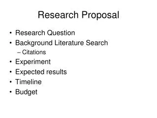
Image format • A-mode (Amplitude) • B-mode (Brightness) B’-mode (1985 O’Donnell) • C-mode (Constant Depth) • D-mode (Depth) • M-mode (Motion) • Color Flow mode • 3-D mode • TissueHarmonic Imaging
Amplitude-mode 250 200 150 100 50 0 0 500 1000 1500 2000 2500 3000 3500 4000
50 45 500 40 1000 35 1500 30 2000 25 20 2500 15 3000 10 3500 5 4000 50 100 150 200 250 300 350 400 Brightness-mode
Depth-mode 40 35 6.93 30 10.01 25 20 depth(mm) 13.09 15 16.17 10 5 19.25 2.5 5 7.5 10 12.5 2.5 5 7.5 10 12.5 position(mm)
20 0 -20 PRI -40 -60 -80 -100 0 50 100 150 200 250 300 350 400 Motion-mode Fast time Slow time
20 40 60 80 100 120 50 100 150 200 250 300 350 400 Motion-mode
3-D mode Surface Rendering Volume Rendering
TissueHarmonic Imaging Fundamental image Harmonic image
MHz MHz LPF Spectrum Spectrum Harmonic Imaging Received Signal Transmit Signal HPF TissueHarmonic Imaging Traditional Filtering Method :
Spectrum Positive pulse MHz MHz SUM MHz Negative pulse Transmit Signal Received Signal TissueHarmonic Imaging Pulse Inversion Technique :
Constant depth-mode • Constant depth scan • Holography • Tomography C-scan
年代/公司別 Acuson ATL Toshiba Siemens GE 3rd Party 5,152,294 1992 1993 核心專利 5,329,929 1994 1995 5,474,073 5,425,365 5,396,890 1996 5,485,842 5,568,812 1997 5,623,930 5,669,385 1998 5,840,034 1999 5,997,480 5,860,924 2000 6,080,108 2001 6,293,914 6,245,017 6,352,509 6,280,387 Patent map
Core patent • PN:5,474,073,ATL (Advanced Technology Laboratories, Inc.),Dec. 1995,being sited by 54 patents • An ultrasonic diagnostic system and scanning technique for producing three dimensional ultrasonic image displays
Core technique • Three dimensional ultrasonic diagnostic image rendering • Combining B-scan & C-scan 2D to 3D scan conversion (ATL & Toshiba) • C-scan method with a linear transducer for measuring the volume flow of fluid in an enclosed structure (Acuson) • Three-dimensional tissue/flow ultrasound imaging system (Siemens)
Research method • Combine several linear scans to a C-scan • Find what spacing is the optimal choice of considering timing and space resolution • Combine 2-D images to depth dependant tomography image or a 3-D image. • Using filters to Volume definition
3-D flow image • Air and Bone • Flow estimation in non image plane (X,Z) • 2-D array or C-scan
Expect results • Tomography and holography presentation. • Find out 3-D flow calculation possibility