Download
1 / 53
540 likes | 554 Views
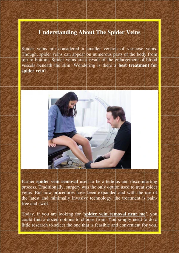
THE VEINS. Introduction The Pulmonary Veins The Systematic Veins. Veins normally accompany arteries and often have similar names. Veins are always larger than the arteries and are sometimes more visible than arteries because they are closer to the skin surface.

E N D
THE VEINS • Introduction • The Pulmonary Veins • The Systematic Veins
Veins normally accompany arteries and often have similar names. Veins are always larger than the arteries and are sometimes more visible than arteries because they are closer to the skin surface. Most veins eventually empty the un-oxygenated blood into the vena cavas.
The Veins General feature • Thin walls, larger lumens, venous valves, venous plexus, venous rete • Two sets • Superficial vein • Deep veins • Special structures: • Sinuses of dura mater • Diploic veins
The Veins Composition • The pulmonary veins • R. & L. superior & inferior pulmonary veins → left atrium • The systemic veins • Superior vena cava system • Inferior vena cava system (hepatic portalsystem ) • Cardiac vein system • →right atrium
The head, neck, upper limb and the thoracic cavity return blood toto the right atrium of the heart via the superior vena cava. The lowerlimb,abdominal and plevis cavity return blood to the right atrium of the heart via the inferior venacava.
Before blood is returned to the heart from the stomach, pancreas, small intestine, and spleen, it goes through the liver for filtration.
This portion of this part of systemic system is known as the hepatic portal system. The gastric vein (stomach), splenicvein (spleen), pancreaticvein (pancreas), and mesentericveins (small intestines) empty into the portal vein that carries the blood to the liver.
In the liver, the portal vein branches into smaller venules and finally into capillary beds. In the capillary beds of the liver, nutrients are exchanged for storage and the blood is purified. The capillaries then join into venules that empty into the hepatic vein, which carries blood to the inferior vena cava.
A. Introduction The Recurrent Factors of the Blood in Veins The venous valves. The dilation of the ventricles of the heart. The negative pressure of the thoracic cavity. The movements of the viscera. The impulses of the artery. the continuous blood flow from capillaries.
B. The Pulmonary Veins Pulmonary Veins 1. Right superior pulmonary v. 2. Right inferior pulmonary v 3. Left superior pulmonary v. 4. Left inferior pulmonary v. the above all left ventricle
C. The Systematic Veins 1. The superior vena caval system 2. The inferior vena caval system
C. The Systematic Veins The superior vena caval system Head & neck internal jugular v. Superior vena cava Brachio- cephalic v. Venous angle Upper limb Subclavian v. Thorax Azygos vein
Veins of head and neck Facial vein • Begins at medial angle of eye (angular vein) • Runs downward and backward through the face, posterior to the facial artery • Below angle of mandible, joins anterior branch of retromandibular vein to form common facial vein, which drains into internal jugular vein
Veins of head and neck Facial vein • Connections with cavernous sinus through the ophthalmic vein , and also through pteygoid venous plexus via the deep facial vein
Veins of head and neck “Danger triangle of the face” • lies between root of nose and two angles of mouth • In this area the facial vein has no valves Connections with cavernous sinus and pteygoid venous plexus
Veins of head and neck Retromandibular v. • Formed by union of superficial temporal and maxillary veins • Divides into an anterior branch that unites with facial vein and a posterior branch that joins posterior auricular vein to become external jugular vein
Veins of head and neck ★External jugular vein • Formed behind angle of mandible by union of posterior auricular, posterior branch of retromandibular and occipital vein • Crossing sternocleidomastoid to enter subclavian vein • Anterior jugular vein • Drains submandibular and anterior neck regions • Descends near midline, runs posterior to sternal end of sternocleidomastoid to drain into external jugular vein or subclavian vein
Veins of head and neck ★ Internal jugular vein • Begin at jugular foramen, descending to join the subclavian vein to form brachiocephalic vein • Lies lateral first to internal and then to common carotid a. within carotid sheath • Chief extracranial tributaries • Common facial vein • Lingual v. • Pharyngeal v. • Superior thyroid v. • Middle thyroid v.
Veins of head and neck Subclavian vein • Continuation of axillary vein at the lateral border of first rib • Joins internal jugular vein to form the brachiocephalic vein. ★Venous angle the junction of the subclavian vein and internal jugular vein
Veins of the Head and Neck Inferior thyroid vein Brachiocephalic vein
Veins of the Upper Limb B. Superficial veins 1. Cephalic v. 2. Basilic v. 3. Median cubital v.
Veins of the Upper Limb 4. Median anterbrachial v. 5. Dorsal venous rete of hand [intravenous drip ] [intravenous injection]
Superficialveins of the upper limb Cephalic vein • Arises from the lateral side of the dorsal venous rete on the back of hand • Winds around the lateral border of the forearm; it then ascends into the cubital fossa and up the front of the arm on the lateral side of the biceps. • It continues up in the deltopectoral groove and then to the infraclavicular fossa, where it pierces clavipectoral fascia to drain into axillary vein.
Superficialveins of the upper limb Basilic vein • Arises from the medial side of the dorsal venous rete of hand • Winds around the medial border of the forearm; it then ascends into the cubital fossa and up the front of the arm on the medial side of the biceps to middle of the arm where it pierces the deep fascia and joins the brachial vein or axillary vein
Superficialveins of the upper limb Median cubital vein • Links cephalic vein and basilic vein in the cubital fossa • It is a frequent site for venipuncture to remove a sample of blood or add fluid to the blood
Variations in the venous pattern of the upper limb Superficial veins are variable and are of significance clinically
Veins of the Upper Limb A. Deep Veins (follow the arteries given the same name:) 1. Subclavian v. 2. Axillary v. 3. Brachial v. 4. Radial v. 5. Ulnar v.
Veins of the Thorax • Parietal Tributaries Anterior Wall of Thorax 1. Thoracoepigastric v. axillary v. 2. Anterior intercostal vv. internal thoracic v. brachiocephalic v.
Veins of the Thorax Posterior Wall of Thorax 1. Post. intercostal vv. left side: accessory hemiazygos v. hemiazygos v. right side:azygos v. sup. vena cava
Veins of the Thorax B. Visceral Tributaries 1. Esophageal v. 2. Bronchial v.
Veins of thorax • Brachiocephalic veinsFormed by union of internal jugular and subclavian veins posterior to the sternoclavicular joint
Veins of thorax • Superior vena cava • Formed by union of right and left brachiocephalic veins behind the right sternocostal synchorndrosis of first rib • Runs vertically down on right of ascending aorta • Joined by azygos vein at level of sternal angle • Enters right atrium at lever of lower border of third right sternocostal joint • Collects blood from veins of upper half of body
Veins of thorax • Azygos vein • Begins as continuation of right ascending lumbar vein • Ascending along the right side of vertebral column • Joins superior vena cava by aching above right lung root at level of T4 to T5 • Receives right posterior intercostals and subcostal veins plus some of bronchial, esophageal and pericardial veins, and hemiazygos vein • Tributaries • Hemiazygos v. • Accessory hemiazygos v.
Veins of thorax Veins of vertebral column • External vertebral venous plexus • Internal vertebral venous plexus
The inferior vena caval system Wall and paired viscera Abdomen Inferior vena cava Unpaired viscera Liver Hepatic v. Pelvis Internal iliac v. Common iliac v. Lower limb External iliac v.
Veins of the Abdomen Unpaired Visceral Tributaries The Hepatic Portal Venous System Digestive apparatus Spleen Inferior vena cava Hepatic poral v. ( Liver Hepatic v)
Veins of the Lower Limb B.Superficial Veins 1. Great saphenous v. → femoral v. 1) superficial medial femoral v. 2) superficial lateral femoral v. 3) superficial iliac circumflex v. 4) superficial epigastric v. 5) external pudendal v.
Veins of the Lower Limb 2. Small saphenous v. popliteal v. 3. Dorsal venous arch of foot
Veins of lower limb • Great saphenous v. • Begins the medial end of dorsal venous arch of foot • Passes anterior to the medial malleolus and ascends on the medial side of the leg, then passes behind the knee and curves forward around the medial side of the thigh • Inclines anteriorly through the thigh to enter the femoral vein through the saphenous hiatus which lies about 3~4 cm below and lateral to the pubic tubercle • Tributaries: • Superficial medial femoral v. • Superficial lateral femoral v. • External pudendal v. • Superficial epigastric v. • Superficial iliac circumflex v.
Superficial epigastric v. Superficial circumflex iliac v. Superficial medial femoral v. Superficial lateral femoral v. Great saphenous v. External pudendal v.
Veins of lower limb Small saphenous v. • Arises from the lateral part of the dorsal venous arch of foot • Ascends behind lateral malleolus and then runs up the midline of the back of the leg • Pierces the deep fascia and enters the popliteal v. • Drains the lateral side of the foot and ankle and the back of the leg.
Veins of the Lower Limb • Deep Veins • External iliac v. 2. Femoral v. 3. Popliteal v. 4. Anterior tibial v. 5. Posterior tibial v.
Veins of pelvis • Internal iliac vein • Parietal tributaries: accompany with arteries • Visceral tributaries • External iliac vein– accompany the artery • Common iliac vein– formed by union of internal and external iliac veins in front of sacroiliac joint, end upon L4~L5 by uniting each other to form inferior vena cava
Veins of the Pelvis A. Parietal Tributaries 1. Superior gluteal v. 2. Inferior gluteal v. 3. Obturator v. 4. Lateral sacral v.
Veins of pelvis Internal iliac vein • Parietal tributaries: accompany with arteries • Visceral tributaries ① Rectal venous plexus ② Vesical venous plexus ③ Uterine venous plexus
Veins of the Pelvis B. Visceral Tributaries 1. Rectal venous plexus →superior rectal vein→inferior mesenteric v. →inferior rectal vein→internal iliac v. →anal vein→internal pudendal v. 2. Vesical venous plexus →vesical v. 3. Uterine venous plexus →uterine v. 4. Veins of perineum internal pudendal v.
Inferior vena cava • Formed by union of two common iliac veins anterior and just to the right of L4~L5 • Ascends on the right side of aorta, pierces vena cava foramen of diaphragm at the level of T8, and drains into the right atrium • Conveys blood from the whole body below the diaphragm to the right atrium
Inferior vena cava Chief tributaries • Parietal • Paired inferior phrenic v. • paired lumbar v. (four) • Visceral • Right and left renal veins • Right suprarenal vein (left drain into left renal vein) • Right testicular or ovarian v. (left drain into left renal vein) • Hepatic veins : right, left and intermediate
Hepatic portal vein General features • Formed behind the neck of pancreas by the union of superior mesenteric vein and splenic vein • Ascends upwards and to the right, posterior to the first part of duodenum and then enters the lesser omentum to the porta hepatis, where it divides into right and left branches • There are no functioning valves in hepatic portal system • Drains blood from gastrointestinal tract from the lower end of oesophagus to the upper end of anal canal, pancreas, gall bladder, bile ducts and spleen