Download
1 / 54
570 likes | 596 Views
Retinoblastoma. Dr Anupam Assistant Professor Ophthalmology. Epidemiology. The most common primary intraocular malignancy of childhood 3% of all childhood cancers The second most common malignant intraocular tumour 1 in 18,000 live births 25-30% cases are bilateral but asymmetric

E N D
Retinoblastoma Dr Anupam Assistant Professor Ophthalmology
Epidemiology • The most common primary intraocular malignancy of childhood • 3% of all childhood cancers • The second most common malignant intraocular tumour • 1 in 18,000 live births • 25-30% cases are bilateral but asymmetric • Survival rates are over 95% in specialized centers but are much lower in the developing world.
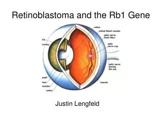
Genetics & Histopathology • RB1 is the tumour suppressor gene in which mutations/deletion predisposes to retinoblastoma. • Tumoursare composed of small basophilic cells (retinoblasts) with large hyperchromatic nuclei and scanty cytoplasm. • Characterized by the formation of structures known as rosettes • Flexner–Wintersteiner, • Homer–Wright and fleurettes
Growth may be endophytic (into the vitreous) with seeding of tumour cells throughout the eye, or • Exophytic (into the subretinal space) leading to retinal detachment, or mixed, or the retina may be diffusely infiltrated.
Optic nerve invasion may occur, with spread of tumour along the subarachnoid space to the brain. • Metastatic spread is to regional nodes, lung, brain and bone.
Types of retinoblastoma 1. Heritable (hereditary, germline) retinoblastoma accounts for 40%. • One of the pair of alleles of RB1 is mutated in all the cells in the body. • When a further mutagenic event (‘second hit’ according to the ‘two-hit’ hypothesis proposed by Knudson) affects the second allele, the cell may then undergo malignant transformation.
Because of the presence of the mutation in all cells, a large majority of these children develop bilateral and multifocal tumours. • Pinealoblastoma (‘trilateral retinoblastoma’, which occurs in up to 10%, usually before the age of 5), • Osteosarcomas, • Soft tissue sarcomas and melanomas
The risk of a second malignancy is about 6% • Increases five-fold if external beam irradiation has been used to treat the original tumour, • The second tumour tends to arise within the irradiated field.
2. Non-heritable (non-hereditary, somatic) retinoblastoma. • 60% Cases • The tumour is unilateral, • Non transmissible and • Does not predispose the patient to second non-ocular cancers.
If a patient has a solitary retinoblastoma and no positive family history, Its very likely that • Non-heritable • The risk in each sibling and the patient’s offspring is about 1%.
Screening of at-risk family members. • Siblings at risk of retinoblastoma should be screened by • Prenatal ultrasonography, • Ophthalmoscopy soon after birth and • Then regularly until the age of 4 or 5 years.
Early diagnosis correlates with a higher chance of preserving vision, salvaging the eye and preserving life. • If a child has heritable retinoblastoma, the risk to siblings is 2% if the parents are healthy, and 40% if a parent is affected. • Parents should also be screened
Clinical features • Bilateral cases present with in 1 yr of age • Unilateral cases present up to 2 yrs of age • Leukocoria (white pupillary reflex) is the commonest presentation (60%) and may first be noticed in family photographs.
Strabismus is the second most common (20%); fundus examination is therefore mandatory in all cases of childhood squint. • Painful red eye with secondary glaucoma • Painful red eye with uveitis • Poor vision. • Inflammation or pseudoinflammation • Nystagmus
Clinical features • Routine examination of a patient known to be at risk. • Orbital inflammation mimicking orbital or preseptal cellulitis may occur with necrotic tumours • Orbital invasion or visible extraocular growth • Metastatic disease involving regional lymph nodes and brain before the detection of ocular involvement is rare.
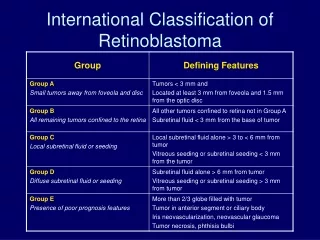
Signs • An intraretinaltumour is a homogeneous, dome-shaped white lesion that becomes irregular, often with white flecks of calcification.
Signs • An endophytictumour projects into the vitreous as a white mass that may ‘seed’ into the gel.
Signs • An exophytictumour forms multilobularsubretinal white masses and causes overlying retinal detachment
Clinical Stages • I. Quiescent stage. • II. Glaucomatous stage. • III. Stage of extraocular extension. • IV. Stage of distant metastasis.
Clinical Stages • I. Quiescent stage. • Lasts for about 6 months to1year. • Leukocoria • Nystagmus • Strabismus • Diminution of vision • Retinal detachment
Clinical Stages • II. Glaucomatous stage. • Pain, redness, watering. • Eyeball is enlarged leading to proptosis. • Conjunctival congesion. • Corneal haze. • Increased intraocular pressure. • Rarely acute iridocyclitis.
Clinical Stages • III. Stage of extraocular extension. • Fungation and involvement of extraocular tissues resulting in marked proptosis
Clinical Stages • IV. Stage of distant metastasis. • 1. Lymphatic spread to preauricular and neighbouring lymph nodes. • 2. Direct extension by continuity to the optic nerve and brain is common. • 3. Metastasis by blood stream involves cranial and other bones.
Investigations • Red reflex testing with a distant direct ophthalmoscope • Examination under anaesthesia • General examination • Tonometry. • Measurement of the corneal diameter • Anterior chamber examination • Ophthalmoscopy, • Cycloplegic refraction.
Investigations • Ultrasonography • Aqueous LDH levels • Wide field photography • CT scan • MRI for optic nerve evaluation • Bone scans and bone marrow aspiration • Genetic study
USG • B Scan displays a caulifiower like mass arising from the retina. • A scan through the mass shows a characteristic V-Y pattern.
Differential diagnosis • Persistent anterior fetal vasculature (persistent hyperplastic primary vitreous) • Coats disease • Retinopathy of prematurity • Toxocariasis • Uveitis • Vitreoretinal dysplasia • Endophthalmitis
Treatment • 1. Tumour destructive therapy. • When tumour is involving less than half of retina and optic nerve is not involved • Chemoreduction followed by local therapy (Cryotherapy, thermochemotherapy or brachytherapy) for large tumours (>12mm in diameter)
Tumour <12 mm in diameter and <8mm in thickness Radiotherapy (external beam radiotherapy or brachytherapy) combined with chemotherapy is recommended for medium size. • Cryotherapy is indicated for a small tumour (<4.5 mm indiameter and <2.5 mm in thickness) located anterior to equator.
Laser photocoagulation is used for a small tumour located posterior to equator <3 mm from fovea. • Thermotherapy with diode laser is used for a small tumour located posterior to equator away from macula
2. Enucleation • Tumour involves more than half of the retina. • Optic nerve is involved. • Glaucoma is present and anterior chamber is involved. • Followed by radiotherapy and chemotherapy if optic nerve is involved. • Intravenous carboplatin, etoposide and vincristine (CEV) are given in three to six cycles according to the grade of retinoblastoma.
Careful review at frequent intervals is generally required following treatment, in order to detect recurrence or the development of a new tumour, particularly in heritable disease.
Palliative therapy • Retinoblastoma with orbital extension, • Retinoblastoma with intracranial extension, and • Retinoblastoma with distant metastasis. • Chemotherapy, • Surgical debulking of the orbit or orbital exentration, and • External beam radiotherapy
Prognosis • If untreated the prognosis is almost always bad and the patient invariably dies. • Rarely, spontaneous regression with resultant cure and shrinkage of the eyeball may occur due to necrosis followed by calcification • Prognosis is fair (survival rate 70-85%) if the eyeball is enucleated before the occurrence of extraocular extension.
MCQs 1. Gene Rb1 responsible for retinoblastoma is located at: • 13q14 • 14q13 • 13p14 • 14p13
MCQs 2. Pathognomic feature of retinoblastoma is: • Necrosis • Calcification • Granulomatous reaction • None
MCQs 3. Characteristic histopathological feature of retinoblastoma is: • Flexner–Wintersteiner rosettes, • Granulomatous reaction • Homer–Wright and fleurettes • None
4. On A scan characteristic pattern of Retinoblastoma is: • Collar stud appearance • V-Y Pattern • Cauliflower pattern • All
5. The most common clinical presentation of retinoblastoma is: • Nystagmus • Leukocoria • Strabismus • Secondary glaucoma
6. Which not a clinical presentation of Retinoblastoma • Nystagmus • Leukocoria • Strabismus • Growth retardation
7. Which not a differential diagnosis for Retinoblastoma • Coat’s disease • Persistent hyperplastic primary vitreous • Endophthalmitis • Central retinal vein occlusion