Download
1 / 71
710 likes | 715 Views
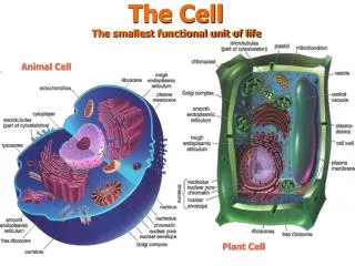
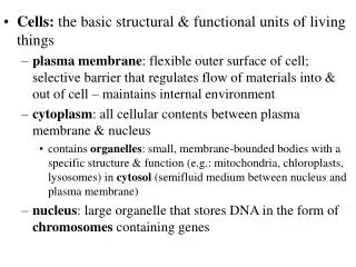
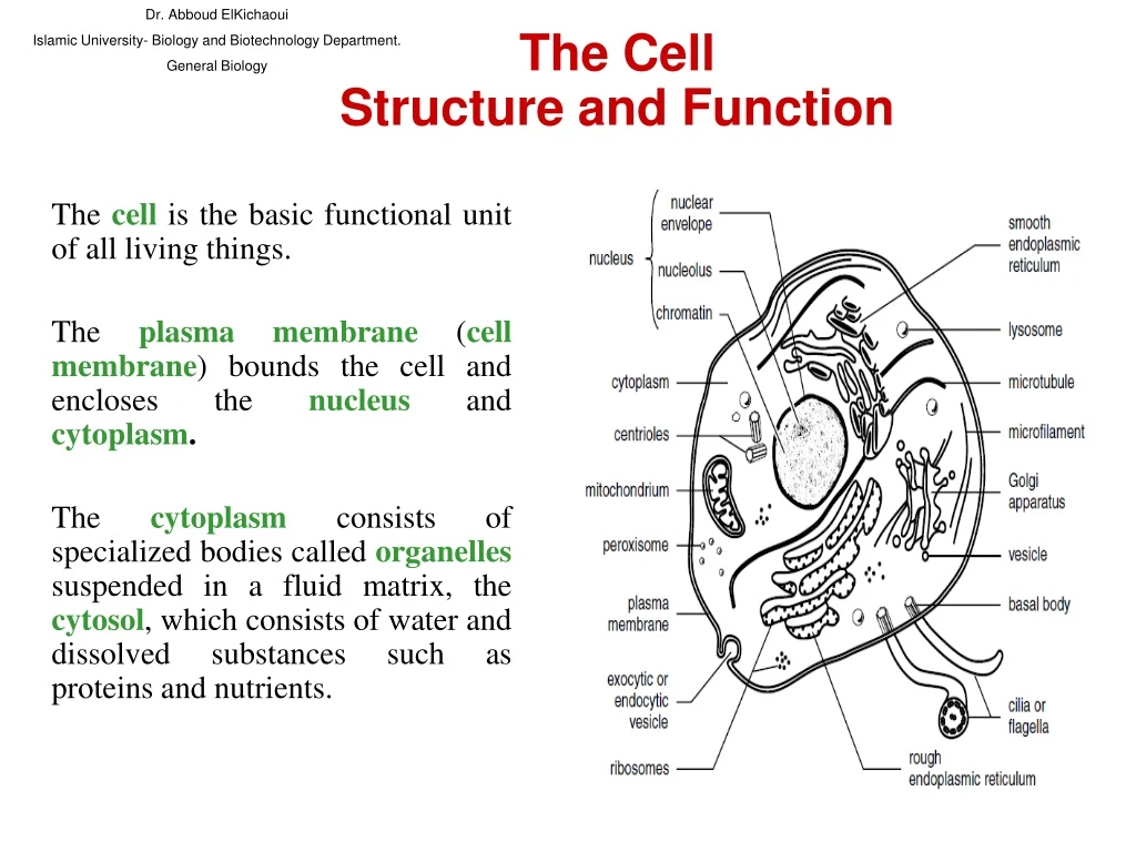
Dr. Abboud ElKichaoui Islamic University- Biology and Biotechnology Department. General Biology. The Cell Structure and Function. The cell is the basic functional unit of all living things. The plasma membrane ( cell membrane ) bounds the cell and encloses the nucleus and cytoplasm .

E N D
Dr. Abboud ElKichaoui Islamic University- Biology and Biotechnology Department. General Biology The Cell Structure and Function The cellis the basic functional unit of all living things. The plasma membrane (cell membrane) bounds the cell and encloses the nucleusand cytoplasm. The cytoplasmconsists of specialized bodies called organellessuspended in a fluid matrix, the cytosol, which consists of water and dissolved substances such as proteins and nutrients.
The plasma membrane The plasma membrane separates internal metabolic events from the external environment and controls the movement of materials into and out of the cell. The plasma membrane is a double phospholipid membrane (lipid bilayer) with the polar hydrophilic heads forming the two outer faces and the nonpolar hydrophobic tails pointing toward the inside of the membrane. Dr. Abboud ElKichaoui Islamic University- Biology and Biotechnology Department. General Biology
Proteins are scattered throughout the flexible phospholipid membrane. • Proteins may attach loosely to the inner or outer surface of the membrane (peripheral proteins), or they may extend into the membrane (integral proteins). Integral proteins may span across the membrane, appearing at both surfaces (transmembrane proteins).
Like phospholipids, integral proteins are amphipathic, with the hydrophobic regions embedded in the membrane and the hydrophilic regions exposed to the aqueous solutions bordering the membrane.
The mosaic nature of scattered proteins within a flexible matrix of phospholipid molecules describes the fluid mosaic model of the cell membrane.
Additional features of the plasma membrane follow: 1. The phospholipid • The phospholipid membrane is selectively permeable • Only small, uncharged, polar molecules (such as H2O and CO2) and hydrophobic molecules (nonpolar molecules like O2 and lipid-soluble molecules such as hydrocarbons) freely pass across the membrane. • In contrast, large polar molecules (such as glucose) and all ions are impermeable.
2. Proteins Proteins in the plasma membrane provide a wide range of functions and include the following: • Channel proteins provide passageways through the membrane for certain hydrophilic (water-soluble) substances such as polar and charged molecules. • Transport proteins spend energy (ATP) to transfer materials across the membrane. When energy is used for this purpose, the materials are said to be activelytransported, and the process is called active transport.
• Recognition proteins distinguish the identity of neighboring cells. These proteins are glycoproteins because they have short polysaccharide chains (oligosaccharides) attached. The oligosaccharide part of the glycoprotein protrudes from the surface of the membrane like an antenna. • Adhesion proteins attach cells to neighboring cells or provide anchors for the internal filaments and tubules that give stability to the cell. oligosaccharides Recognition proteins Adhesion proteins
Receptor proteins provide binding sites for hormones or other trigger molecules. In response to the hormone or trigger molecule, a specific cell response is activated. • Electron transfer proteins are involved in transferring electrons from one molecule to another during chemical reactions.
Cholesterol Cholesterol Cholesterol Cholesterol 3. Cholesterol Cholesterol molecules distributed throughout the phospholipid bilayer provide some rigidity to the plasma membranes of animal cells. In plant cells, related substances (sterols) provide a similar function. CYTOPLASM Cholesterol helps maintain the fluidity of the membrane by preventing the phospholipids from packing too tightly.
4- The glycocalyx The glycocalyx is a carbohydrate “coat” covering the outer face of the plasma membrane. It consists of various oligosaccharides that are attached to membrane phospholipids (glycolipids) and proteins (such as the glycoproteins of recognition proteins). The glycocalyx provides markers for cell-cell recognition.
Organelles Organelles are bodies within the cytoplasm that serve to physically separate the various metabolic reactions that occur within cells Dr. Abboud ElKichaoui Islamic University- Biology and Biotechnology Department. General Biology
Dr. Abboud ElKichaoui Islamic University- Biology and Biotechnology Department. General Biology
1. The nucleus The nucleus is bounded by the nuclear envelope, a phospholipid bilayer similar to the plasma membrane. The nucleus contains DNA (deoxyribonucleic acid), the hereditary information of the cell. Normally, the DNA is spread out within the nucleus as a threadlike matrix called chromatin. Dr. Abboud ElKichaoui Islamic University- Biology and Biotechnology Department. General Biology
Nucleus cont…. When the cell begins to divide, the chromatin condenses into rod-shaped bodies called chromosomes, each of which, before dividing, is made up of two long DNA molecules and various histone (protein) molecules. The histones serve to organize the lengthy DNA, coiling it into bundles called nucleosomes. Dr. Abboud ElKichaoui Islamic University- Biology and Biotechnology Department. General Biology
Nucleus cont…... Also visible within the nucleus are one or more nucleoli, concentrations of DNA in the process of manufacturing the components of ribosomes. The nucleus also serves as the site for the separation of chromosomes during cell division.
2- Ribosome • Ribosome subunits are manufactured in the nucleus and consist of RNA molecules and proteins. • The two subunits, labeled 60S and 40S, move across the nuclear envelope and into the cytoplasm where they are assembled into a single 80S ribosome. • (An S value, or Svedberg unite expresses how readily a product forms a sediment in a centrifuge, with larger values representing larger and heavier products). Large subunit Small subunit Dr. Abboud ElKichaoui Islamic University- Biology and Biotechnology Department. General Biology Svedberg unite = S = The sedimentation rate for a particle
In the cytoplasm, ribosomes assist in the assembly of amino acids into proteins.
3. The endoplasmic reticulum (ER) • The endoplasmic reticulum, or ER, consists of stacks of flattened sacs involved in the production of various materials. • In cross section, they appear as a series of maze-like channels, often closely associated with the nucleus. Dr. Abboud ElKichaoui Islamic University- Biology and Biotechnology Department. General Biology
Rough ER When ribosomes are present, the ER (called rough ER) creates glycoproteins by attaching polysaccharide groups to polypeptides as they are assembled by the ribosomes. Dr. Abboud ElKichaoui Islamic University- Biology and Biotechnology Department -General Biology
Smooth ER, • Smooth ER without ribosomes, is responsible for various activities, including the synthesis of lipids and hormones, especially in cells that produce these substances for export from the cell. • In liver cells, smooth ER is involved in the breakdown of toxins, drugs, and toxic by-products from cellular reactions. Dr. Abboud ElKichaoui Islamic University- Biology and Biotechnology Department. General Biology
4. Golgi apparatus (Golgi complex or Golgi body) A Golgi apparatus (Golgi complex or Golgi body) is a group of flattened sacs arranged like a stack of bowls. They function to modify and package proteins and lipids into vesicles, small, spherically shaped sacs that bud from the outside surface of the Golgi apparatus. Dr. Abboud ElKichaoui Islamic University- Biology and Biotechnology Department. General Biology
Transport vesiclefrom Golgi Transport vesiclefrom ER Rough ER Plasmamembrane Vacuole Nucleus Lysosome Golgiapparatus Smooth ER Nuclearenvelope • Vesicles often migrate to and merge with the plasma membrane, releasing their contents to the outside of the cell.
5. Lysosomes Lysosomes are vesicles from a Golgi apparatus that contain digestive enzymes. Dr. Abboud ElKichaoui Islamic University- Biology and Biotechnology Department. General Biology
They break down food, cellular debris, and foreign invaders such as bacteria. Lysosomes do not occur in plant cells. Dr. Abboud ElKichaoui Islamic University- Biology and Biotechnology Department. General Biology Formation and Function of Lysosomes Flash for Lysosomes formation.
6. Peroxisomes Peroxisomes Peroxisomes are organelles that break down various substances. During the breakdown process, O2 combines with hydrogen to form toxic hydrogen peroxide (H2O2), which in turn is converted to H2O. Peroxisomes are common in liver and kidney cells where they break down toxic substances and in photosynthesizing plant cells. Peroxisomes transfer hydrogen from various substances to oxygen Dr. Abboud ElKichaoui Islamic University- Biology and Biotechnology Department. General Biology
7. Mitochondria Mitochondria carry out aerobic respiration, a process in which energy (in the form of ATP) is obtained from carbohydrates. Dr. Abboud ElKichaoui Islamic University- Biology and Biotechnology Department. General Biology
8. Chloroplasts Chloroplasts carry out photosynthesis, the plant process of incorporating energy from sunlight into carbohydrates. Dr. Abboud ElKichaoui Islamic University- Biology and Biotechnology Department. General Biology
9. Microtubules, intermediate filaments, and microfilamentsMicrotubules, Microtubules, intermediate filaments, and microfilaments are three protein fibers of decreasing diameter, respectively. All are involved in establishing the shape of or in coordinating movements of the cytoskeleton, the internal structure of the cytoplasm. Dr. Abboud ElKichaoui Islamic University- Biology and Biotechnology Department. General Biology
A- Microtubules Microtubules are made of the protein tubulin and provide support and motility for cellular activities. They are found in the spindle apparatus (which guides the movement of chromosomes during cell division), and in flagella and cilia (described in the following section), structures that project from the plasma membrane to provide motility to the cell. spindle apparatus (which guides the movement of chromosomes Dr. Abboud ElKichaoui Islamic University- Biology and Biotechnology Department. General Biology
B- Intermediate filaments Intermediate filaments provide support for maintaining the shape of the cell. Dr. Abboud ElKichaoui Islamic University- Biology and Biotechnology Department. General Biology
C- Microfilaments Microfilaments are made of the protein actin and are involved in cell motility. They are found in muscle cells and in cells that move by changing shape, such as phagocytes(white blood cells that wander throughout the body attacking bacteria and other foreign invaders). microfilamentsinmuscle phagocytes Dr. Abboud ElKichaoui Islamic University- Biology and Biotechnology Department. General Biology
10. Flagella and cilia Flagella and cilia are structures that protrude from the cell membrane and make wavelike movements. Flagella and cilia are classified by their lengths and by their numbers per cell: flagella are long and few; cilia are short and many. A single flagellum propels sperm, while the numerous cilia that line the respiratory tract sweep away debris. Dr. Abboud ElKichaoui Islamic University- Biology and Biotechnology Department. General Biology
Flagella and cilia cont….. Structurally, both flagella and cilia consist of microtubules arranged in a “9 + 2” array–nine pairs (doublets) of microtubules arranged in a circle surrounding a pair of microtubules Basal bodyA cellular organelle associated with the formation of cilia and flagella and similar to the centriole in structure.
FLAGELLUM Electron micrograph of sections: Outer microtubule doublet Plasmamembrane Flagellum Centralmicrotubules Outer microtubule doublet Plasmamembrane Basal body Basal body(structurally identical to centriole)
11. Centrioles and basal bodies Centrioles and basal bodies act as microtubule organizing centers (MTOCs). A pair of centrioles (enclosed in a centrosome) located outside the nuclear envelope gives rise to the microtubules that make up the spindle apparatus used during cell division. Early mitoticspindle Centrosomes(with centriole pairs) Centrosome Chromatin Plasmamembrane Spindlemicrotubules
Basal bodies of centriole are at the base of each flagellum and cilium and appear to organize their development. • Both centrioles and basal bodies are made up of nine triplets arranged in a circle.
Life cycle of Ferns السَّرْخَس Life cycle of Mosses حَزازُ • Plant cells lack centrioles and only “lower” plants (such as mosses and ferns) with motile sperm have flagella and basal bodies.
Dr. Abboud ElKichaoui Islamic University- Biology and Biotechnology Department. General Biology
12. Cell walls Cell walls are found in plants, fungi, protests, and bacteria. They develop outside the plasma membrane and provide support for the cell. In plants, the cell wall consists mainly of cellulose, a polysaccharide made from β-glucose. The cell walls of fungi are usually made of cellulose or chitin. Chitin is a modified polysaccharide differing from cellulose in that one of the hydroxyl groups is replaced by a group containing nitrogen. Dr. Abboud ElKichaoui Islamic University- Biology and Biotechnology Department. General Biology
13. Vacuoles and vesicles Vacuoles and vesicles are fluid-filled, membrane-bound bodies. • Transport vesiclesmove materials between organelles or between organelles and the plasma membrane. • Food vacuolesare temporary receptacles of nutrients. Food vacuoles often merge with lysosomes, whose digestive enzymes break down the food. • Storage vacuolesin plants store starch, pigments, and toxic substances (nicotine, for example).
Central vacuolesare large bodies occupying most of the interior of certain plant cells. When fully filled, they exert turgor, or pressure, on the cell walls, thus maintaining rigidity in the cell. They also store nutrients and carry out functions otherwise assumed by lysosomes in animal cells.
Contractile vacuolesare specialized organelles in single-celled organisms that collect and pump excess water out of the cell.
14. Cell junctions Cell junctions serve to anchor cells to one another or to provide a passageway for cellular exchange. They include the following: • Desmosomes • Tight junctions • Gap junctions • Plasmodesmata Dr. Abboud ElKichaoui Islamic University- Biology and Biotechnology Department. General Biology
• Desmosomes Desmosomes are protein attachments between adjacent animal cells. Inside the plasma membrane, a desmosome bears a disk-shaped structure from which protein fibers extend into the cytoplasm. Desmosomes act like spot weldsبقعة اللحام to hold together tissues that undergo considerable stress (such as skin or heart muscle). Dr. Abboud ElKichaoui Islamic University- Biology and Biotechnology Department. General Biology
• Tight junctions Tight junctions are tightly stitched seams between animal cells. The junction completely encircles each cell, preventing the movement of material between the cells. Tight junctions are characteristic of cells lining the digestive tract where materials are required to pass through cells (rather than intercellular spaces) to penetrate the blood stream. Dr. Abboud ElKichaoui Islamic University- Biology and Biotechnology Department. General Biology
• Gap junctions Gap junctions are narrow tunnels between animal cells that consist of proteins called connexons. The proteins prevent the cytoplasms of each cell from mixing, but allow the passage of ions and small molecules. In this manner, gap junctions allow communication between cells through the exchange of materials or through the transmission of electrical impulses. Dr. Abboud ElKichaoui Islamic University- Biology and Biotechnology Department. General Biology
• Plasmodesmata (singular, Plasmodesmata) are narrow channels between plant cells. A narrow tube of endoplasmic reticulum, called a desmotubule, surrounded by cytoplasm and the plasma membrane, passes through the channel. Material exchange through a Plasmodesmata apparently occurs through the cytoplasm surrounding the desmotubule. Dr. Abboud ElKichaoui Islamic University- Biology and Biotechnology Department. General Biology