Download
1 / 24
260 likes | 531 Views
Human Reproductive System Review. Borrowed from Mrs. Degl. Male Reproductive System. Male Reproductive System. External Structures. Internal Structures. Prostate : exocrine gland of male reproductive system Vas Deferens : tubes connecting epididymis to ejaculatory ducts

E N D
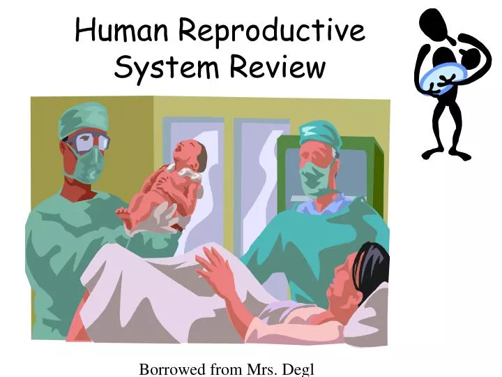
Human Reproductive System Review Borrowed from Mrs. Degl
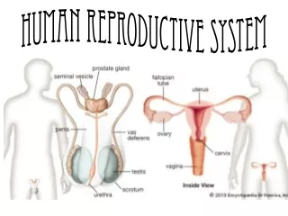
Male Reproductive System External Structures Internal Structures Prostate: exocrine gland of male reproductive system Vas Deferens: tubes connecting epididymis to ejaculatory ducts Epididymis: organ where sperm matures Testicles: organ where sperm is created Urethra: tube that connects bladder to outside of body • Penis: external male sex organ Scrotum: sac of skin and muscle containing testicles
Sperm Formation • MEIOSIS forms sperm in gametes • Sperm forms in testes • Scrotum keeps cooler than rest • Semen is sperm plus protecting fluid • Semen leaves testes through the vas deferens (sperm ducts) to the urethra before it exits.
Secondary Sexual Characteristics - Male • Produced by testosterone • Deeper voice • Chest and facial hair • Lengthen bones • Increased size of testes for sperm production
Female Reproductive System Mrs. Degl
Female Reproductive System Internal Structures External Structures Mons Pubis: soft mound of flesh above genitals Labia: lip-like structures on the outside of the vagina • Vagina: tract from uterus to exterior • Hymen: mucous membrane around vaginal opening • Cervix: lower, narrow portion of uterus • Uterus: pear-shaped organ containing growing fetus • Fallopian Tubes: pathway for egg travel during ovulation • Fimbria-tissue near the ovaries • Ovaries: egg-producing organs
Fimbria The fimbria is a fringe of tissue near the ovary leading to the Fallopian tubes. When ovulation is about to occur, the sex hormones activate the fimbria, causing it to hit the ovary in a gentle, sweeping motion.
Ovary Ovaries are part of the vertebrae female reproductive system. Normally, a female will have two ovaries, each performing two major functions: producing eggs and secreting hormones. Ovaries in females are homologous to testes in males. The term gonads refer to the ovaries in females and testes in males.
Uterus The main function of the uterus is to accept a fertilized ovum, which becomes implanted into the endometrium, and derives nourishment from blood vessels which develop exclusively for this purpose. The fertilized ovum becomes an embryo, develops into a fetus and gestates until childbirth. Due to anatomical barriers such as the pelvis, the uterus is pushed partially into the abdomen due to its expansion during pregnancy. Even in pregnancy the mass of a human uterus amounts to only about a kilogram.
Cervix The opening from the vagina into the womb allows menstrual blood exit and sperm in.
Vagina During live birth, the vagina provides the route to deliver the fetus from the uterus to its independent life outside the body of the mother. During birth, the vagina is often referred to as the birth canal.
Secondary Sexual Characteristics - Female • Induced by increased LH, FSH, estrogen, and progesterone hormone levels • Pubic hair • Widen pelvis • Enlarge mammary tissue (breasts) • Begin menstrual cycles
Ovum Formation • MEIOSIS forms the eggs (ova) • Eggs are formed before birth • 1 egg per month is matured and released from ovaries, most of the time • Eggs travel through the fallopian tube where they may become fertilized
Fertilization • Occurs in upper 1/3 of Fallopian tube • Only 1 sperm can fertilize an egg • Fertilized egg = zygote • An average woman is pregnant (gestational period) for 10 whole months. That is 40 weeks. Babies can survive if they are born earlier, but they may have complications due to being premature. • At 36 weeks a baby is considered term
Implantation • Fertilized eggs are implanted into thick walls of uterus • Chorion membranes dig into uterus to form placenta • Zygote grows into an embryo • Embryo gets air and nutrients through the umbilical cord • Once pregnant, progesterone levels stay high in mom • Mom’s uterus grows with the baby
First Trimester • Pregnancy is counted in weeks, lasting 40 weeks from the first day of your last period. So you are actually only preparing for pregnancy during those first two weeks, until ovulation. For two more weeks many women do not know that they are pregnant, even though they may be hoping that they conceived this month. • About the time your next period is due is when pregnancy tests begin to pick up the first traces of hCG in your urine or blood. • The picture to the right is 5 days after conception.
1st Trimester = 1st 12 weeks • Heart develops first • Neural tube develops • All body systems appear by Week 8 – Now a Fetus
Second Trimester • Not only have most women ceased being nauseated, many feel a burst of energy and report feeling the best that they've ever felt. • The baby is finishing it's development and at the end of this trimester she or he will begin to put more weight on. Major organ systems are functioning and fetal movement can be felt by mom and outside parties by the end of the second trimester. Some women will have ultrasound screening around 20 weeks gestation. About 50% of families will choose to find out the sex of their baby at this point as well.
2nd Trimester = up to 24 weeks • Most growth • Looks more like a baby • Some preemies survive at this stage Video Clip
Third Trimester • Baby is getting bigger and loving life in the womb. Many babies will start to settle into a head down position, beginning as early as the 28th week. • About 3-4% of all babies will remain in the breech position at the end of pregnancy. This final trimester is really a time for finishing touches like lung maturity and layers of brown fat to help keep your baby warm on the outside.
3rd Trimester= Up to 40 weeks • More growth • Kicking, rolling, stretching • Eyes open – Week 32 • Lungs mature • Rotates to head-down position, unless baby is breech
Birth • Labor • Uterine contractions begin • Cervix dilates to 10 cm. • Birth • Uterus pushes baby through vaginal canal • Placenta delivered after baby • Cesarean Section (c-section) is a surgery that cuts through the uterus to deliver the baby if it cannot be born vaginally