Download
1 / 50
500 likes | 733 Views
Role of MRI in TOF follow-up. TOF symposium October 25, 2013 Dr Edythe Tham. Outline. Quantification of RV size & function Quantification of pulmonary regurgitation Pulmonary stenosis Branch pulmonary arteries Conduits and artificial valves. Goals of cardiac MRI.

E N D
Role of MRI in TOF follow-up TOF symposium October 25, 2013 Dr Edythe Tham
Outline Quantification of RV size & function Quantification of pulmonary regurgitation Pulmonary stenosis Branch pulmonary arteries Conduits and artificial valves
Goals of cardiac MRI • Quantification of RV & LV volumes and function (RVEF) • Quantification of pulmonary regurgitant fraction (RF) • Anatomy of the RVOT & branch pulmonary arteries (and aorta) • Assessment of myocardial fibrosis
Pulmonary regurgitation Transannular patch
Pulmonary Regurgitation Region of interest
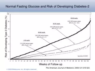
Criteria for pulmonary valve replacement • RVEDV >170 ml/m2 • RVESV > 85 ml/m2 • RVEF < 45% • Regurgitant Fraction >30% Therrien et al, AJC 2005
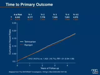
Relationship between RV volume and pulmonary regurgitation Samyn et al, JMRI 2007
Relationship between RV ESV & RVEF Geva et al, JACC 2004
17 year female, S/P TAP RVEDVi 111 ml/m2 RVESVi 56 ml/m2 RVEF 50% LVEF 60%
Regurgitant Fraction 43%
11 year female with TOF/PAS/P RV-PA conduit RVEDVi 178 ml/m2 RVESVi 150 ml/m2 RVEF 16% LVEF 28%
Regurgitant fraction 57% Peak velocity 2 m/s = Peak gradient 16 mmHg
10 year female S/P TAP Mixed disease – Mild PS: 20 mmHg Moderal PR: 34%
Magnetic Resonance Angiography Branch pulmonary arteries
21 year male S/P TOF repair RPA 56%: LPA 44% Mild proximal LPA stenosis, PG 25 mmHg
18 year old S/P TOF repair – bilateral branch PA stenosis RPA 75%: LPA 25% Peak gradients: RPA: 38 mmHg LPA: 29 mmHg
12 year female with branch PA stenosis From MRI RPA 82%: LPA 18%
Left pulmonary artery Right pulmonary artery
Artifact from prosthetic valve • 12 year female • Prosthetic pulmonary valve • Melody valve
38 year male S/P 29 mm Hancock valve RVEDVi 170 ml/m2 RVESVi 98 ml/m2 RVEF 42% RF 20% Peak velocity 3 m/s = PG 36 mmHg
Indications for cardiac MRI • Baseline post-TOF repair at 7-10 years (no sedation required) • Follow up every 1-3 years depending on clinical status • Yearly MRI if: symptomatic or evidence of RV dysfunction
Cardiac MRI: Disadvantages Not portable Contraindications: pacemaker/AICD Affected by metallic artifacts eg prosthetic valves, stents
Advantages of MRI No radiation Does not require sedation in older children Independent of acoustic windows Capability for 3D reconstruction Quantifies ventricular function Flow quantification