Download
1 / 33

E N D
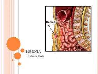
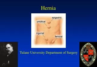
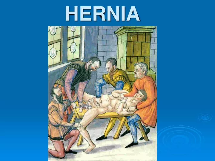
HERNIA • Hernia of the abdominal wall or external hernia is such surgical disease, which is characterized by outlet of the visceral organs from the place of their physiological placement through the natural channels or defects of the abdominal and pelvic wall. In such case all visceral organs covered by parietal peritoneum and skin are not damaged. • Internal hernia is such disease, visceral organs hit the peritoneum pouch. It formed in the place of natural peritoneum fold or recess and generally kept in the abdominal cavity.
The Walls Of The Inguinal Canal ANTERIOR WALL • laterally - muscles fibers of the external oblique • medially - aponeurosis of the external oblique • most medially there is not wall but instead there is a deficiency called the superficial inguinal ring. SUPERIOR -- arching fibers of the internal oblique and sometimes transverse abdominis. These fibers start anterior and lateral, pass over the spermatic cord and the medially forms part of the posterior wall of the canal. POSTERIOR -- lateral the posterior wall is deficient at the deep inguinal ring. Medially the posterior wall is made up of the fused aponeuroses of the internal oblique and transverse abdominis, called the conjoined tendon X. INFERIOR (or floor) -- inguinal ligament. Medially, some of the fibers of the inguinal ligament curve under the spermatic cord and fasten into the pectineal line of the pubis, this is the lacunar ligament which forms part of the floor of the inguinal canal.
ETIOLOGY AND PATHOGENESIS • Hernias are divided into two main groups: congenital and acquired. The main reason of congenital hernias is malformation. Thus, inguinal hernia arose in case of noclosure of the process of peritoneum, which passes by inguinal channel during descending the testis. On such hernias testis is located in the hernia pouch. Acquired inguinal hernia has hernia pouch and testis located outside it.
PATHOMORPHOLOGY • Each abdominal hernia consists of hernia gate, hernia sac and hernia contents. Hernia sac forms by outpouching of parietal peritoneum and can contain small intestine and omentum. Sometimes it containes other organs: large intestine, urinary bladder, ovary, and appendix. • The main parts of the hernia pouch are neck, body and fundus.
CLASSIFICATION • 1) Depends on anatomical localization: inguinal (indirect and direct), midline hernia, omphalocele, femoral hernia, lumbar hernia, sciatic hernia, (enterischiocele), lateral hernia, ischiorectal hernia (perineocele). • 2) depends on etiology: congenital (herniae conqenitae) and acquired (herniae acguisitae). • 3) Depends on clinical presentations: complete and incomplete, reducible and nonreducible, traumatic and postoperative, complicated and noncomplicated.
Clinical variants and complications • Inguinal hernias is developed in two ways: through the internal (middle) inguinal cavity and external (lateral). In the first case formed direct in other - indirect inguinal hernia. • Indirect hernias could be congenital and acquired. Direct hernias are only acquired and occur in older patients. • There are two main signs, which differentiate direct and indirect hernias. Direct hernia is always located medially from a. epigastrica inf. Indirect hernia is always located laterally from a. epigastrica inf. The other sign is: direct hernia located medially from deferent duct, indirect hernia located inside it.
DIAGNOSIS PROGRAM • Anamnesis and physical examination. • Digital investigation of the hernia channel. • Sonography of the hernia pouch. • Common blood analysis. • Common urine analysis.
Choice of treatment method • Bassini repair. After extraction of the hernia sac, we are taking spermatic duct on holders. Between the borders of transverse muscle, internal oblique muscle, transverse fascia and inguinal ligament interrupted sutures placed. Except that, couples sutures placed between border of abdominal rectus muscle sheath and pubic bone periosteum. In such way, inguinal space closured and posterior wall strengthened. Spermatic duct placed on the new-formed posterior wall of the inguinal channel. Over the spermatic duct aponeurosis restored by interrupted sutures.
Choice of treatment method • Girard in such kind of the operations propose to attach the edges of the internal oblique muscle and transversal muscle to the inguinal ligament over the spermatic duct. The aponeurosis of the external oblique muscle sutured by second layer of the suture. Excess of the aponeurosis is fixed to the muscle in the form of duplication
Choice of treatment method • Spasokukotskyy proposed to catch the edges of the internal oblique muscle and transversal muscle with aponeurosis of the external oblique muscles by single-layer interrupted suture
Choice of treatment method • Martynov proposed the fixation to the Poupart's ligament only internal edge of the external oblique muscle aponeurosis without muscles. External edge of the aponeurosis sutured over internal in the form of duplication
Choice of treatment method • Kimbarovskyy, based on the principles of joining similar tissues, proposed special suture: Sutures placed on 1 cm from the edge of the external oblique abdominal muscle aponeurosis, grasped the part of the internal oblique and transversal muscle. After that, aponeurosis is sutured one more time from behind to the front and attached to the Poupart’s ligament
Choice of treatment method • Postempskyy proposed the deaf closing of inguinal interval with the латеральним moving of spermatic duct. The plastic narrowing of internal inguinal ring of to 0,8 cm is the important moment of this modification. On occasion, when internal and external inguinal rings are in one plane, a spermatic duct is displaced inlateral direction by transversal incision of the oblique and transversal muscles. Then edge of the vagina of direct muscle and aponeurosis of the internal and transversal muscles is fixed to the Couper’s ligament
Treatment of the femoral hernia • The Bassini method is attributed to “femoral”. It is performed from a cut, that passes under inguinal fold. After removal of hernia sack a hernia gate is liquidated by suturing of inguinal to the pectineal ligament
Treatment of the femoral hernia • The Rudgi-Parlavecho Method. A cut passes parallel to the inguinal fold and higher it (the same as at inguinal hernia). A hernia sack is removed. After that the edges of the transversal and internal oblique muscles and inguinal ligament sutured to the periosteum of pubic bone.
Treatment of umbilical hernia • The Lexer operation is most widespread. It performed by imposition of sutures on an umbilical ring
Treatment of umbilical hernia • After the Meyo method defect of anterior abdominal wall in the umbilical ring is sutured by U-shaped stitches in transversal direction
Treatment of umbilical hernia • Sapezhko proposed to form duplication of the abdominal white line by stitches in longitudinal direction.
INCARCERATED HERNIA • Incarcerated hernia is sudden pressing of hernia contents in a hernia orifice. Incarceration is the most frequent and most dangerous complication of hernia diseases.
Etiology and pathogenesis • At the elastic incarceration, after increasing intraabdominal pressure, one or a few organs relocated from an abdominal cavity to the hernia sack, where it is compressed with following ischemia and necrosis in the area of hernia gate. • At the fecal incarceration in the intestinal loop which is in a hernia sack, plenty of excrement passed quickly. Proximal part of loop is overfilled, and distal is compressed in a hernia gate.
Classification of the incarcerated hernia • complete • Incomplete • partial (the Richter’s hernia) • retrograde • without the destructive changes of hernia contents • with the phlegmon of hernia sack
Clinical variants • Retrograde incarceration • Parietal incarceration (the Richter’s hernia) • The Littre’s hernia • Incarceration at sliding hernia
Diagnosis program • Anamnesis examination. • Physical examination. • Blood analysis and urine analysis. • Digital investigation of the rectum. • Survey X-Ray of abdominal cavity organs.