Download
1 / 1
10 likes | 108 Views
Genetically Engineered Materials Science & Engineering Center Mehment Sarikaya, University of Washington, DMR 0520567. Protein-Driven Synthesis of Transition Metal-Doped ZnS Immuno-Quantum Dots.

E N D
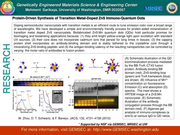
Genetically Engineered Materials Science & Engineering Center Mehment Sarikaya, University of Washington, DMR 0520567 Protein-Driven Synthesis of Transition Metal-Doped ZnS Immuno-Quantum Dots Doping semiconductor nanocrystals with transition metals is an efficient route to tune emission color over a broad range of wavelengths. We have developed a simple and environmentally friendly process for protein-aided mineralization of transition metal doped ZnS nanocrystals. Biofabricated ZnS:Mn quantum dots (QDs) hold particular promise for bioimaging and biosensing applications because: (1) they emit bright yellow-orange light upon excitation with standard UV sources; (2) their core does not incorporate cadmium ions that persist for long times in tissues; (3) the capping protein shell incorporates an antibody-binding domain and is stably tethered to the crystalline core through a mineralizing ZnS binding peptide; and (4) the antigen-binding valency of the resulting nanoparticles can be controlled by varying the molar ratio of antibodies to fusion protein. (A) Schematic illustration of the QD biomineralization process mediated by the BB-TrxA::CT43 fusion protein. Antibody-binding BB domain (red), ZnS-binding loop (green) and TrxA framework (blue) are shown. (B) Influence of Mn2+ concentration on fluorescence. Emission (C) and absorption (D) spectra . The inset shows a HRTEM image of a ZnS:Mn nanocrystal. (E) Schematic illustration of the antibody conjugation process through the BB domain (red). (F) Agarose gel analysis of immuno-complexes (a and b) at various IgG to QD ratios. RESEARCH W. Zhou, D. T. Schwartz, & F. Baneyx. JACS, 132, 4731–4738 (2010). * Supported by NSF via GEMSEC, MRSEC at UW For more information, visit GEMSEC at: http://www.GEMSEC.washington.edu