Download
1 / 46
460 likes | 687 Views
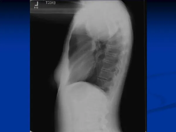
A 19-YO Woman with Huge Mediastinal Mass. She had history of chronic cough and night sweat for 1 month. She had no swelling of face nor upper extremities. Left axillary and left lower cervival lymph node were palpated afterward. Chest film showed large mediastinal mass. CT chest and abdomen

E N D
A 19-YO Woman with Huge Mediastinal Mass • She had history of chronic cough and night sweat for 1 month. She had no swelling of face nor upper extremities. • Left axillary and left lower cervival lymph node were palpated afterward. • Chest film showed large mediastinal mass. • CT chest and abdomen • A large lobulated anterior mediastinal mass (4.5x10x6.5 cm) involving prevascular space of superior mediastinum and surrounding SVC, brachiocephalic vessels, ascending aorta, aortic arch MPA, left main pulmonary artery without obstruction. Multiple small paraaortic LN above IMA origin • Atelectasis and obstructive pneumonia at lingular segment of LUL and bronchopneumonia at aterior segment of LUL
Echocardiography • Good LV contraction, LVEF 65.1%, no LA dilatation, no significant valvular dysfunction • No abnormal wall motion. No intracardiac shunt, no intracardiac mass, normal pericardium, no pericardial effusion
Lymph node biopsy, left cervical group • Diffuse large B-cell lymphoma, anaplastic variant • Some resembling Hodgkin or Reed-Sternberg cells • Lymphoma cells mark distinctly with CD20, not CD3 or EMA. Some mark with CD30 • Bone marrow biopsy • Negative for lymphoma cells • High LDH 1037 U/L (N 225-450), ECOG 2
Diffuse Large B-cell Lymphoma: At Least Three Disease Primary Mediastinal B Cell Lymphoma (PMBL) Activated B Cell-like (ABC DLBCL) Germinal Center B Cell-like (GCB DLBCL) Cell of origin Germinal center B cell ? Post-germinal Center B cell ? Thymic B cell Oncogenic Mechanisms • BCL-2 • Translocation • C-rel • amplification Constitutive activation Of NF-kB Chr. 9q24 Amplification PDL2 Clinical outcome Favorable 59% 5-yr survival Poor 30% 5-yr survival Favorable 64% 5-yr survival
Primary Mediastinal B-Cell Lymphoma (PMBL) • Large cells with polymorphic nuclei that have an abundant rim of clear cytoplasm. Fibrosis commonly results in compartmentalization of the neoplastic cells • Immunophenotyping demonstrates the presence of B-cell antigens in all cases (CD19, CD20, CD22 and CD79a). • Bcl-2 is expressed in 80% of cases, • CD10 is infrequently expressed and CD21 is negative. Surface immunoglobulin (sIg) expression is absent, • CD30 staining is common, but weak. • In contrast to HL, the transcription factors PAX5, BOB.1, Oct-2 and PU.1 are always expressed and CD15 is negative.
Comparison of DLBCL and PMLBCL DLBCLPMLBCL Median age (years) 5535 Nodal/extranodal presentation 65%/35%0%/100% Sex distribution (M:F) 1:11:4 Stage I-II/III-IV 40%/60%80%/20% Bulky disease 30%60%-70% Haematologica 2008;93:1364-71.
PMBL vs HL vs MGZL Dunleavy K, et al. Gray zone lymphoma; better treated like Hodgkin lymphoma or mediastinal large B-cell lymphoma. Curr Hematol Malig Rep 2012;7:241-7.
Diagnosis and Treatment • Diffuse large B-cell lymphoma, primary mediastinal B-cell lymphoma stage IIIBX • IPI = 3 (LDH, stage, ECOG); Bulky • Treatment • R-CHOP-21 x 8 cycles • After CR, autologous stem cell transplantation was done without prior radiotherapy
Interim PET after 2nd R-CHOP • A soft tissue density mass at left anterosuperior mediastinum, 4.5x6.0 cm at prevascular, paraaortic, AP window, lower paratracheal to left hilar region; no cervical lymphadenopathy • No FDG avidity at soft tissue density mass. Mild FDG avidity at left hilar node (residual tumor)
CT whole body after 4th R-CHOP • Decreased size of soft tissue mass at anterior mediastinum, 3.7x1.9x3 cm. No pericardial effusion • Decreased size of multiple lymph nodes at left gastric, EG junction, paraaortic, aortocaval, celiac region
PET/CT after 8th R-CHOP • No pathological size of lymph nodes in head and neck regions, normal orbits and paranasal sinuses • No pulmonary mass or nodule. An ill-defined isodensity mass in anterosuperior mediastinum (5x2.8 cm) occupying in prevascular space, paraaortic and AP window • No demonstrable mass or cyst or fluid in abdominal and pelvic cavity • No demonstrable bony destruction • IMP: complete response, no active lymphoma
Haematologica 2008;93:1364-71. • The recommended first-line therapy is chemotherapy and radiotherapy (grade B). An anthracycline-based chemotherapy with CHOP, MACOP-B or VACOP-B is recommended (grade B) • Patients with an inadequate early response should be candidates for early intensification with high-dose chemotherapy (grade C) • Patients with refractory or relapsed disease should undergo rescue programs including intensive, non-cross-resistant debulking treatment followed, in chemosensitive patients, by high-dose chemotherapy and ABMT (grade B).
Overall survival by chemotherapy subtype in the IELSG study of 426 patients with primary mediastinal large B-cell lymphoma (PMBL) 100% HDS/ABMT, n = 44 80 3rd generation regimens, n = 277 60 40 CHOP, n = 105 P < .0001 426 pts 20 0 2 4 6 10 14 16 18 8 12 years Haematologica 2002;87:1258-126
The Therapeutic Outcome with the Inclusion of Radiation Therapy Haematologica 2002;87:1258-64.
Multivariate analysis of poor prognostic factors influence OS P-value Exp (B) 95% Cl Increasing age 0.0002 1.02 1.01-1.03 Male sex 0.02 1.49 1.05-2.12 Poor performance status 0.001 0.51 0.34-0.77 Advanced stage 0.004 0.57 0.39-0.83 Induction chemotherapy 0.0002 0.49 0.34-0.71
R-CHOP RT vs CHOP RT Vassilakopoulos TP, et al. Rituximab, cyclophosphamide, doxorubicin, vincristine, and prednisone with or without radiotherapy in primary mediastinal large B-cell lymphoma: the emerging standard of care. The Oncologist 2012;17:139-49.
R-CHOP RT vs CHOP RT Vassilakopoulos TP, et al. Rituximab, cyclophosphamide, doxorubicin, vincristine, and prednisone with or without radiotherapy in primary mediastinal large B-cell lymphoma: the emerging standard of care. The Oncologist 2012;17:139-49.
Comparative Outcome of 76 PMBL with R-CHOP RT and 45 Historical Control with CHOP RT Vassilakopoulos TP, et al. Rituximab, cyclophosphamide, doxorubicin, vincristine, and prednisone with or without radiotherapy in primary mediastinal large B-cell lymphoma: the emerging standard of care. The Oncologist 2012;17:139-49.
Early Stage Patients All Patients
Front-line ASCT in PMBL OS PFS DFS Rodriguez J, et al. Primary mediastinal large cell lymphoma (PMBL): frontline treatment with autologous stem cell transplantation (ASCT). The GEL-TAMO experience. Hematol Oncol 2008;26:171-8.
A 30-YR Female with Bilateral Cervical Lymphadenopathy • She had chronic intermittent fever with night sweats for 3 months. Bilateral enlarged cervical lymph nodes were palpated. • Physical examination revealed moderate anemia without jaundice, bilateral cervical and supraclavicular lymphadenopathy, and palpable splenomegaly. • Cervical lymph node biopsy revealed classical Hodgkin lymphoma, nodular sclerosis type.
CT whole body • Multiple matted LN at bilateral supraclavicular regions, right upper paratracheal, paraesophageal, subcarina, intraabdominal cavity, bilateral iliac regions • Hepatomegaly and splenomegaly with multiple splenic lymphoma nodules
Initial Laboratory Results • CBC : Hb 8.6 g/dl, WBC 4880/mm3 (N 75, band 15, L 6, M 2), platelet 295,000/mm3 • ESR 75 mm/h • LDH 964 U/L (N 225-450 U/L) • Albumin 3.2 g/dl, Ca 8.5mg/dl • Bone marrow study • Multifocal marrow necrosis with diffuse myelofibrosis with abnormal medium to large mononuclear cells marked with CD30+, CD20-, CD3-, ALK-, CD15-
Diagnosis • Classical Hodgkin’s lymphoma, nodular sclerosis • Stage IVBS • Advanced Stage with 4 risk factors • Hb < 10.5 g/dl • Stage IV • Albumin < 4 g/dl • Lymphocyte < 8%
GHSG, German Hodgkin Study Group EORTC, European Organisation for Research and Treatment of Cancer GELA, Groupe d’Etude des Lymphomes de l’adulte
Advanced Stage HL • Associated with failure rate 30-40% with anthracycline polychemotherapy • Treatment • Evaluation of regimens comprising multi-agent chemotherapy • Multiple consolidative strategies • Improved disease control
Higher Intensive regimens to increase efficacy • BEACOPP-based regimen (GHSG) • standardBEACOPP • Escalated BEACOPP • BEACOPP-14 • > 2000 patients treated: esc BEACOPP • CR: >90% (RT: 15-65%) • FFTF: 82-88%, 4-10 yr follow up • OS: 86-90%, 4-10 yr follow up • MDS/AML: 0.9% • Significant more hematological and infectious toxicity, secondary leukemia/MDS, infertility
Hodgkin Lymphoma Advanced Stages 1.0 BEACOPP escalated BEACOPP base 0.8 C/ABVD 0.6 Probability Only alkylating agents (1965) 0.4 No treatment (1940) 0.2 0.0 0 1 2 3 4 5 6 7 8 9 10 Overall Survival (y)
Other intensive regimen • Standford V regimen and RT to any bulky disease • Consolidation treatment • Consolidation RT • Patient with poor risk or residual lesions • Less toxic than ASCT • Supported by EORTC (MOPP/ABV IFRT) in PR cases • Not recommended in PET-CR patients (GELA H89, GHSG H15) • Autologous stem cell transplant (ASCT) • Only in very high risk patient • Not recommended in 1st CR patient
Risk Adapted Treatment • Assessing chemosensitivity by interim PET • Prognostic value of PET after 2 cycles of chemotherapy (PET2) • PET2 – negative better FFS • High negative predictive value for disease progression • Low positive predictive value • Ongoing trial • 2xABVD PET2+ BEACOPP-14 or EB; PET2- ABVD or ABV (UK RATHL) • 2xEB PET2+6xEB rituximab; PET2- 2x vs 6xEB (GHSG HD18)
Gallamini A, et al. Early interim 2-[18F] Fluoro-2-deoxy-D-glucose positron emission tomography is prognostically superior to international prognostic score in advanced-stage Hodgkin’s lymphoma: a report from a joint Italian-Danish study. JCO 2007;25(24):3746-52.
BEACOPPesc vs ABVD in advanced HL • 3 Italian cooperative group • Michelangelo Foundation • The Gruppo Italiano di Terapie Innovative nei Linfomi (GITIL) • The Intergruppo Italiano Linfomi (IIL) • Unfavorable and advanced HL patients (331 patients) • 6x ABVD • 4x BEACOPPesc/ 4x BEACOPPstd • For CR and VGPR pt 30 G IFRT • For <CR, relapse salvage with ifosfamide-based and HDT/ASCT with BEAM Viviani S, et al. NEJM 2011;365(3):203-12.
Better initial control in BEACOPP No difference in long-term clinical outcome Viviani S, et al. NEJM 2011;365(3):203-12.
esc BEACOPP vs ABVD in early unfavorable and advanced stage HL • 4 published trials (2868 adult patients) • GHSG HD9 and HD14 from Germany • HD2000 and GSM-HD from Italy • PFS significantly longer for escBEACOPP • OS, not statistically significant • More toxicity in escBEACOPP than ABVD • Hematological, infection, AML/MDS • No differences in 2nd cancer, TRM or infertility • 16-60 adult patients with unfavorable/ advanced HL benefited from escBEACOPP for PFS, no difference in OS Bauer K, et al. Cochrane Database of Systematic Reviews 2011, Issue 8.
Bauer K, et al. Cochrane Database of Systematic Reviews 2011, Issue 8.
Case continued • The patient was treated with ABVD x4 • CT whole body after 4x ABVD • Much improvement of multiple lymph nodes and masses • Small cervical, surpraclavicular nodes < 1 cm • No mediastinal mass • Multiple paraaortic node 1-1.7 cm • Normal live and spleen • No pelvic mass, no ascites
Before After ABVDx4
Case continued • Much improvement after 4 cycles of ABVD • Another 2 cycles of ABVD was added • PET/CT after completion of 6th ABVD was done • No hypermetabolic or enlarged lymph node in neck, chest and intraabdominal regions and no abnormal uptake in the bony structure that indicated no evidence of active lymphoma • Suggestive of hypermetabolic intramural myoma at posterior fundus