Download
1 / 3
0 likes | 7 Views
GAS AND VAPOUR ANALYSIS BY MOHAMED ANWE RIFKY FROM ZAGAZIG MEDICAL SCHOOL

E N D
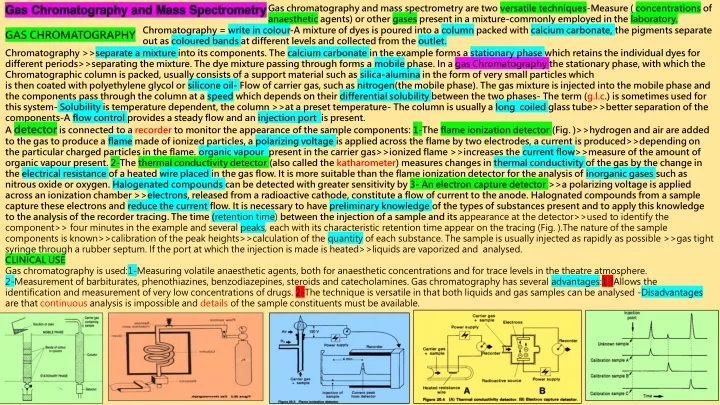
Gas Chromatography and Mass Spectrometry Gas chromatography and mass spectrometry are two versatile techniques-Measure ( concentrations of anaesthetic agents) or other gases present in a mixture-commonly employed in the laboratory. Chromatography = write in colour-A mixture of dyes is poured into a column packed with calcium carbonate, the pigments separate out as coloured bands at different levels and collected from the outlet. GAS CHROMATOGRAPHY Chromatography >>separate a mixture into its components. The calcium carbonate in the example forms a stationary phase which retains the individual dyes for different periods>>separating the mixture. The dye mixture passing through forms a mobile phase. In a gas Chromatography the stationary phase, with which the Chromatographic column is packed, usually consists of a support material such as silica-alumina in the form of very small particles which is then coated with polyethylene glycol or silicone oil- Flow of carrier gas, such as nitrogen(the mobile phase). The gas mixture is injected into the mobile phase and the components pass through the column at a speed which depends on their differential solubility between the two phases- The term (g.l.c.) is sometimes used for this system- Solubility is temperature dependent, the column >>at a preset temperature- The column is usually a long coiled glass tube>>better separation of the components-A flow control provides a steady flow and an injection port is present. A detector is connected to a recorder to monitor the appearance of the sample components: 1-The flame ionization detector (Fig. )>>hydrogen and air are added to the gas to produce a flame made of ionized particles, a polarizing voltage is applied across the flame by two electrodes, a current is produced>>depending on the particular charged particles in the flame. organic vapourpresent in the carrier gas>>ionized flame >>increases the current flow>>measure of the amount of organic vapour present. 2-The thermal conductivity detector (also called the katharometer) measures changes in thermal conductivity of the gas by the change in the electrical resistance of a heated wire placed in the gas flow. It is more suitable than the flame ionization detector for the analysis of inorganic gases such as nitrous oxide or oxygen. Halogenated compounds can be detected with greater sensitivity by 3- An electron capture detector >>a polarizing voltage is applied across an ionization chamber >>electrons, released from a radioactive cathode, constitute a flow of current to the anode. Halognated compounds from a sample capture these electrons and reduce the current flow. It is necessary to have preliminary knowledge of the types of substances present and to apply this knowledge to the analysis of the recorder tracing. The time (retention time) between the injection of a sample and its appearance at the detector>>used to identify the component>> four minutes in the example and several peaks, each with its characteristic retention time appear on the tracing (Fig. ).The nature of the sample components is known>>calibration of the peak heights>>calculation of the quantity of each substance. The sample is usually injected as rapidly as possible >>gas tight syringe through a rubber septum. If the port at which the injection is made is heated>>liquids are vaporized and analysed. CLINICAL USE Gas chromatography is used:1-Measuring volatile anaesthetic agents, both for anaesthetic concentrations and for trace levels in the theatre atmosphere. 2-Measurement of barbiturates, phenothiazines, benzodiazepines, steroids and catecholamines. Gas chromatography has several advantages:1-lAllows the identification and measurement of very low concentrations of drugs. 2-The technique is versatile in that both liquids and gas samples can be analysed -Disadvantages are that continuous analysis is impossible and details of the sample constituents must be available.
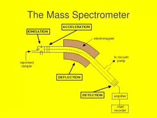
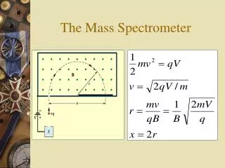
THE MASS SPECTROMETER FURTHER TECHNIQUES OF GAS AND VAPOUR ANALYSIS The sample is drawn through a tube into the sample chamber of a mass spectrometer by a pump. A molecular leak permits a few molecules of the sample >>an ionization chamber , bombarded by a beam of electrons passing from the hot cathode to the anode. When the molecules of the gas are hit by the electrons,>>become charged ions which are then accelerated out of the chamber in a narrow beam by means of the acceleration and focusing plates. The stream of ions >>a strong magnetic field. The charged ions are deflected in an arc by the magnetic field, the amount of deflection depending on their mass (lighter ions being deflected most). In the (Fig.) four different ion streams are shown, only one of which passes through a small slit to be picked up by a detector, and this signal can be amplified and displayed. By varying the voltage on the acceleration and focusing plates, the position and speed of the beam may be altered and streams of ions of different masses detected. Another method of picking out specific streams of ions >>useing four electrically charged rods in place of the magnetic field. The potentials on the rods are varied so that the ions oscillate between them as they travel. It can be arranged so that only ions of a specific mass are able to travel the length of the rods without being removed from the stream. (quadrupole mass spectrometer).Many molecules fragment during the ionization process>>carbon dioxide, carbon monoxide and oxygen ions, having mass numbers of 44, 28 and 16 respectively. Some of the ions have the same mass number >>ionized fragment of the compound of interest which has a different mass number from any of the other ions present>>nitrous oxide in the presence of carbon dioxide, as both have the same mass number of 44>> nitric oxide fragment with a mass number of 30 can be identified .The mass spectrometer is a versatile instrument >>measure many different gases and compounds, with a rapid response time of less than 100 ms, used for the breath by breath analysis of expired air. and can analyse very small samples . OTHER TECHNIQUES OF VAPOUR ANALYSIS Gas Chromatograph and mass spectrometer are highly versatile but, bulky and less convenient for use in areas outside the laboratory. Anaesthetists often use alternative techniques in such areas. 1-The solubility of anaesthetic agents in oil >> an electric potential is applied across a crystal of quartz >> contracts slightly (piezoelectric effect)>>crystal can be made to oscillate at its resonant frequency with a thin oily coating being applied. Anaesthetic agents dissolve in the oily coating and alter the resonant frequency of the crystal. From (Henry’s law) thequantity of vapour which dissolves is proportional to the partial pressure of the vapour. 2- Vapour concentration can also be measured by the infrared analyser(refractometer). 3-Measurement of the velocity of ultrasound in a gas mixture and the application of Raman spectrometry.
FURTHER TECHNIQUES OF GAS AND VAPOUR ANALYSIS THE RAMAN EFFECT A-Interaction of the electromagnetic waves with molecules: 1-The radiation is scattered (E.of scattered radiation = incidence radiation). 2-The molecule absorb all of the radiation (CO2 absorbing infrared radiation). 3-Partial absorbtion between both (Raman effect). B-In Ramant effect>> E. absorbed of particular length>>specific type of bond between the atoms in the molecule. As E. of radiation is prop. to the freq.>> transfer of the radiation and the molecule >> change in wave length. C-It is a weak effect>>intense light as Laser is used, and the axis of the detector is perpendicular to the axis of light source. D-Its principle: 1-A helium -neon Laser between two mirrors. 2-Set of eight detectors ( only 2 in the fig.).Each detector comprises a filter >> R.of specific wave length and a photodetector (incident R.>> electrical signal>>processing).Filters made to pass Raman R. specific for O2,N2 CO2 N2O,the C-H bond and 3 volatile anaesth. agents. 3-water is removed from the sample>> dry. E-The signal from the photodetector measures radiation from the C-H bond >> conc. of anaesth. agents (usually one at a time ).The signal obtained is stronger than others. However, radiation from individual concentration agents are still used. F-Intensity of Laser>>degrading anaesth. agents>> pumped out the window (affect measuring ). RAMAN IS FROM INDIA.WINNING A NOBEL PRIZE 17-GAS AND VAPOUR ANALYSIS: A- HOW DOES THE GAS CHROMATOGRAPHY WORK ? B- GIVE A FULL ACCOUNT ON THE MASS SPECTROMETER. C- GIVE A FULL ACCONT ON RAMAN EFFECT. Having great diversity or variety = Versatile A team of researchers from Massachusetts Institute of Technology (MIT) and Massachusetts General Hospital (MGH) hopes to improve those numbers with a new sensor using Raman spectroscopy that can be embedded into an epidural needle, helping anaesthesia doctors guide the needle to the correct location. Raman spectroscopy (/ˈrɑːmən/) (named after physicist C. V. Raman) is a spectroscopic technique typically used to determine vibrational modes of molecules,