Download

1 / 9
120 likes | 366 Views
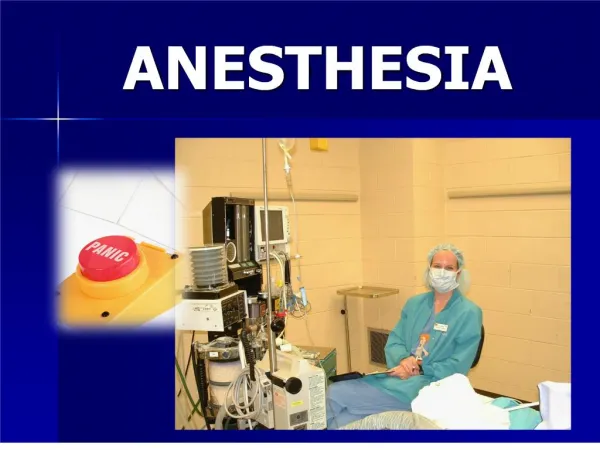
Anesthesia . Preparation . Removal of food and water is recommended for 12 hours prior to surgery

E N D
Preparation Removal of food and water is recommended for 12 hours prior to surgery Supplemental heat is also used to maintain the patient at approximately 85 degrees F. It is also important to keep this temperature consistent throughout the anesthetic induction, the surgical procedure, and the recovery phase. Water circulating heating pad
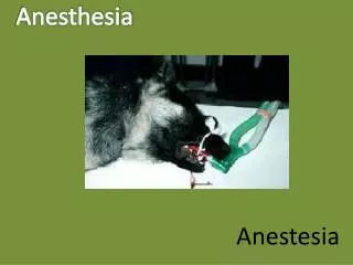
Anesthesia machine • Isoflurane and, more recently, sevoflurane are the inhalant anesthetic agents of choice for use in birds • Oxygen
Induction • Mask induction is common, starting at 5% for less than a minute, decrease to 3% then maintain in 1.5% • A high flow rate of 2l/min of O2 is utilized due to the large amount of dead space w/in the mask • Monitoring depth of anesthesia in birds is different than in dogs and cats since palpebral reflex, toe pinch, and jaw tone are unreliable • If a bird is shivering, the plane of anesthesia is very light
ET • Once intubated, the O2 flow a rate is set at app. 1L/min • The eyes of the bird should be lubricated ASAP • Since birds posses complete tracheal rings, which are not distensible, it is necessary to place uncuffed ET tubes • Inflation of cuffed ET tubes can cause pressure necrosis and sloughing of the tracheal mucosa
Air sac tube • Due to the uniqure respiratory anatomy of birds in which O2 exchange occurs on both inspiration and expiration an ET tube can be placed into the caudal thoracic air sac through the lateral body wall to provide not only O2 but inhalant gas anesthesia as well
Air sac • Air sac tubes are instrumental in ER cases of tracheal occlusion, or when sx of the trachea or head area is necessary • To place an air sac tube, make a 2mm skin incision in the area of the lateral body wall that is just caudal to the last rib and just ventral to the lateral process of the relatively thin abdominal musculature taking care not to traumatize underlying organs • An ET tube is placed through the open jaws of the hemostat and sutured in place
Monitoring • A Doppler, placed on the medial metatarsal artery or radial artery, and a pulse ox, placed over the femur, foot, toe, or humerus are helpful, but nothing replaces vigilantly and constantly observing breathing, and periodically ausculting the HR w/ a stethoscope
Recovery • It is fast • Ensure to maintain optimal temperature of the particular species for faster drug metabolism (and recovery) • Provide a secure and clear airway • Provide adequate analgesia : Butorphanol is administered IM in painful procedures