Download
1 / 30
300 likes | 456 Views
Structure and gating mechanism of the acetylcholine receptor pore. Acetylcholine receptor. Important in transmitting messages from neuron to neuron Is a excitatory neurotransmitter Acetylcholine is stored in vesicles at the end of a neuron

E N D
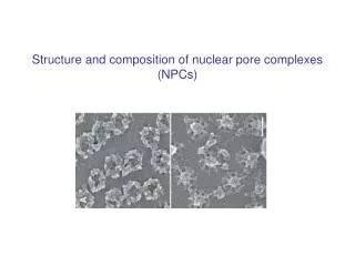
Structure and gating mechanism of the acetylcholine receptor pore
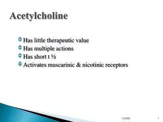
Acetylcholine receptor • Important in transmitting messages from neuron to neuron • Is a excitatory neurotransmitter • Acetylcholine is stored in vesicles at the end of a neuron • When the right signal comes along, it causes the release of acetylcholine into the synaptic cleft • Here it binds to the acetylcholine receptor and which will allow for further transmission of the signal
Propagation of signal from neuron to neuron via neurotransmitters
Acetylcholine receptor • Classic example of a ligand gated channel • So it is a ion channel that is ligand gated • It ultimately leads to an influx of positive ions
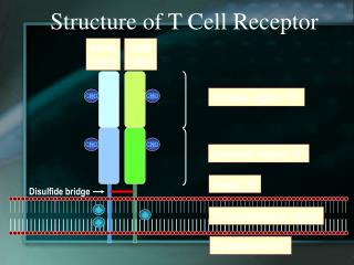
Ach Receptor Structure • 5 transmembrane polypeptides and they are 2 alpha, 1 beta, 1 delta, and 1 gamma • Each subunit is made up of 4 transmembrane alpha helices (helical bundle). They are M1-M4
Each helices has features • M2 is mostly amphipathic while M1, M3, and M4 are mostly hydrophobic • M2 has polar side chains for the most part in the middle of the pore and hydrophobic backing up against the back of the pore
Inside the channel • In the middle of the channel there are 5 leucine sidechains and these 5 leucine sidechains come 1 from each M2 helix • There function is to protrude into the channel and block it • This is when the channel is in its closed conformation and inactivated
The activated Ach Receptor • Bulky hydrophobic leucines are removed from channel core • This occurs when Ach binds to the receptor • Binding causes conformational change, twisting of the M2 helices and the leucines twist out of the way
The Activated Ach Receptor continued • The M2 helices now instead of having bulky hydrophobic residues now have polar residues facing inward and ions can go down the channel
Ach Receptor Summary • Bulky hydrophobic leucine side chains of M2 helices close the channel • Upon activation from Ach binding, the leucine side chains twist out of the way and now the polar hydrophilic side chains and now positive ions can rush through.
The Paper about Ach Receptor • Used liquid high helium temperatures • Resolved protein structure at 4A • Proposed a mechanism of how the pore opens and closes in response to presence or absence of Ach
Method • Obtained crystals from Torpedo marmorata membranes • Crystals at first did not yield high amplitudes so they adjusted their procedure using Fourier transforms to enhance signal • Used electron imaging to obtain their structures
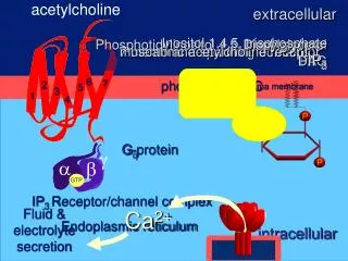
Signal Transduction • When cells convert an extracellular signal to an intracellular signal that alter the behavior of the target cell
Ionotropic Channel Very fast Metabotropic Second messenger slower Recptors
Receptors • Ion-channel-linked (AchR) ionotropic • G-protein linked metabotropic • Enzyme-linked metabotropic involves the covalent modification of P to serine threonine or tyrosine
G protein receptor • 7 transmembrane helices • Links outside to inside of cell
G protein • Made up of three subunits, alpha, beta, and gamma ( A heterotrimeric protein) • The alpha subunit is referred to as the key player in relaying the signal
Activated G protein leads to the exchange of GDP (inactive form) to GTP (active form) • Then this activated subunit will then go and activate another protein
Alpha Subunit • GTP will bind at a site on this subunit and activate it Gfrom Transducin
Pic of Beta Subunit Beta Subunit • Built from 7 WD repeats ( motifs that are built from tryptophan(W) and aspartate(D) • Beta Propellar formed by seven subunits made of Beta Sheets
Putting it all together • G does not interact with Gbut the are linked by the Gsubunit • GGinteract • Forms functional signaling unit
GEF GDP GTP GAP
Ras protein • Monomeric • Is active when it has GTP bound • Play a role in cell growth and differentiation • GTP linked to Ras via Mg2+