Download
1 / 16
160 likes | 277 Views
M icroPET : Radiotracer I maging of Rodents and Non- H uman Primates. Alexander K. Converse, PhD University of Wisconsin–Madison Waisman Center - Brain Imaging Core Town Hall - Thursday 2 February 2012. Radioactive Decay & Positron – Electron Annihilation. P ositron E mission T omography.

E N D
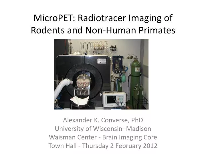
MicroPET: Radiotracer Imaging of Rodents and Non-Human Primates Alexander K. Converse, PhD University of Wisconsin–Madison Waisman Center - Brain Imaging Core Town Hall - Thursday 2 February 2012
3 uL 6 uL 2 uL + MR Animal Brain PET Scanners 3 uL
PHARMACOKINETIC MODELING Fig. 25: QUANTIFICATION: Logan determination of binding of a dopamine D2 tracer5 Fig. 23. PARAMETER ESTIMATION: Time activity curves from a multiple injection study of a dopamine D2 receptor ligand in rhesus15 Fig. 24 DOPAMINE RELEASE: Time activity curves of a dopamine D2 receptor tracer in response to amphetamine5 Fig. 26 BLOOD VOLUME: Carbon monoxide imaging of hemoglobin in rat12
INFLAMMATION Fig. 20 MULTIPLE SCLEROSIS: Microglial cell activation in white matter in response to zymosan in rat11 Fig. 19 ASTHMA: Glucose metabolism in inflammation in a rat lung10
PAIN Fig. 22 AVIAN VETERINARY ANALGESIA: Increased glucose metabolism in response to experimental arthritis in parrot6 Fig. 21 OPIOID RECEPTORS: Kappa opioid receptor availability in parrot brain6
PARKINSON’S DISEASE Fig. 16 NEURODEGENERATION: Rat model of striatal neurodegneration for pre- (left) and post- (right) stem cell treatment (first image from the microPET P4, 2002) Fig. 15 STEM CELL THERAPY: Dopamine synthesis in a rhesus model pre- and post- unilateral lesion13 Fig. 17 GENE THERAPY: Dopamine synthesis pre- and post- lentiviral delivery of GDNF in a unilateral lesion rhesus model8 Fig. 18 L-DOPA: Recovery of AAAD activity in a rhesus model7
MOOD DISORDERS Fig. 7 CHILDHOOD ANXIETY: Dopamine D2 receptors in rhesus1 Fig. 8 CHILDHOOD ANXIETY: Glucose metabolism in rhesus9 Fig. 9 SEXUAL BEHAVIOR: Glucose metabolism in female marmosets
ADDICTIVE BEHAVIORS Fig. 12 PRENATAL ALCOHOL EXPOSURE: Image of dopamine transporter in rhesus and time activity curves4 Fig. 10 PRENATAL ALCOHOL EXPOSURE: glucose metabolism alterations due to a reversal task in rhesus3 Fig. 11 PRENATAL ALCOHOL EXPOSURE: Serotonin 1A receptors in rhesus2 Fig. 13 AMPHETAMINE: Blood flow alteration correlated with dopamine release in rhesus5 Fig. 14: EATING BEHAVIORS Dopamine D2 receptor response to deep brain stimulation in rhesus14
Design Logistics Radiochemistry Scanner Analysis PET
Alex Converse Director, MicroPET Imaging Waisman Center Brain Imaging Core akconverse@wisc.edu